Potassium »
PDB 3ukm-3vrs »
3vjf »
Potassium in PDB 3vjf: Crystal Structure of De Novo 4-Helix Bundle Protein WA20
Protein crystallography data
The structure of Crystal Structure of De Novo 4-Helix Bundle Protein WA20, PDB code: 3vjf
was solved by
R.Arai,
A.Kimura,
N.Kobayashi,
K.Matsuo,
T.Sato,
A.F.Wang,
J.M.Platt,
L.H.Bradley,
M.H.Hecht,
with X-Ray Crystallography technique. A brief refinement statistics is given in the table below:
| Resolution Low / High (Å) | 50.00 / 2.20 |
| Space group | P 21 21 2 |
| Cell size a, b, c (Å), α, β, γ (°) | 65.950, 102.858, 31.344, 90.00, 90.00, 90.00 |
| R / Rfree (%) | 23.3 / 25.5 |
Potassium Binding Sites:
The binding sites of Potassium atom in the Crystal Structure of De Novo 4-Helix Bundle Protein WA20
(pdb code 3vjf). This binding sites where shown within
5.0 Angstroms radius around Potassium atom.
In total only one binding site of Potassium was determined in the Crystal Structure of De Novo 4-Helix Bundle Protein WA20, PDB code: 3vjf:
In total only one binding site of Potassium was determined in the Crystal Structure of De Novo 4-Helix Bundle Protein WA20, PDB code: 3vjf:
Potassium binding site 1 out of 1 in 3vjf
Go back to
Potassium binding site 1 out
of 1 in the Crystal Structure of De Novo 4-Helix Bundle Protein WA20
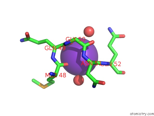
Mono view
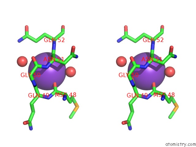
Stereo pair view
Mono view
Stereo pair view
A full contact list of Potassium with other atoms in the K binding
site number 1 of Crystal Structure of De Novo 4-Helix Bundle Protein WA20 within 5.0Å range:
|
Reference:
R.Arai,
N.Kobayashi,
A.Kimura,
T.Sato,
K.Matsuo,
A.F.Wang,
J.M.Platt,
L.H.Bradley,
M.H.Hecht.
Domain-Swapped Dimeric Structure of A Stable and Functional De Novo Four-Helix Bundle Protein, WA20 J.Phys.Chem.B V. 116 6789 2012.
ISSN: ISSN 1089-5647
PubMed: 22397676
DOI: 10.1021/JP212438H
Page generated: Sat Aug 9 06:05:26 2025
ISSN: ISSN 1089-5647
PubMed: 22397676
DOI: 10.1021/JP212438H
Last articles
Zn in 9QM9Zn in 9S44
Zn in 9OFE
Zn in 9OFC
Zn in 9OFD
Zn in 9OF1
Zn in 9OFB
Zn in 9N0J
Zn in 9M5X
Zn in 9LGI