Potassium »
PDB 3ukm-3vrs »
3v7q »
Potassium in PDB 3v7q: Crystal Structure of B. Subtilis Ylxq at 1.55 A Resolution
Protein crystallography data
The structure of Crystal Structure of B. Subtilis Ylxq at 1.55 A Resolution, PDB code: 3v7q
was solved by
N.J.Baird,
J.Zhang,
T.Hamma,
A.R.Ferre-D'amare,
with X-Ray Crystallography technique. A brief refinement statistics is given in the table below:
| Resolution Low / High (Å) | 19.97 / 1.55 |
| Space group | P 21 21 21 |
| Cell size a, b, c (Å), α, β, γ (°) | 50.407, 61.291, 94.160, 90.00, 90.00, 90.00 |
| R / Rfree (%) | 20 / 22.9 |
Potassium Binding Sites:
The binding sites of Potassium atom in the Crystal Structure of B. Subtilis Ylxq at 1.55 A Resolution
(pdb code 3v7q). This binding sites where shown within
5.0 Angstroms radius around Potassium atom.
In total 2 binding sites of Potassium where determined in the Crystal Structure of B. Subtilis Ylxq at 1.55 A Resolution, PDB code: 3v7q:
Jump to Potassium binding site number: 1; 2;
In total 2 binding sites of Potassium where determined in the Crystal Structure of B. Subtilis Ylxq at 1.55 A Resolution, PDB code: 3v7q:
Jump to Potassium binding site number: 1; 2;
Potassium binding site 1 out of 2 in 3v7q
Go back to
Potassium binding site 1 out
of 2 in the Crystal Structure of B. Subtilis Ylxq at 1.55 A Resolution
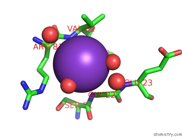
Mono view
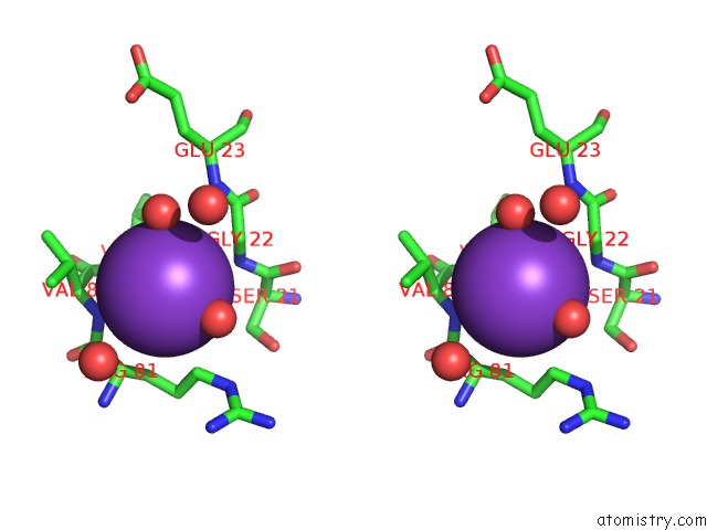
Stereo pair view
Mono view
Stereo pair view
A full contact list of Potassium with other atoms in the K binding
site number 1 of Crystal Structure of B. Subtilis Ylxq at 1.55 A Resolution within 5.0Å range:
|
Potassium binding site 2 out of 2 in 3v7q
Go back to
Potassium binding site 2 out
of 2 in the Crystal Structure of B. Subtilis Ylxq at 1.55 A Resolution
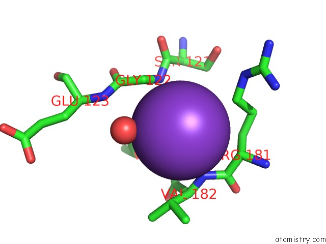
Mono view
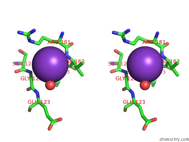
Stereo pair view
Mono view
Stereo pair view
A full contact list of Potassium with other atoms in the K binding
site number 2 of Crystal Structure of B. Subtilis Ylxq at 1.55 A Resolution within 5.0Å range:
|
Reference:
N.J.Baird,
J.Zhang,
T.Hamma,
A.R.Ferre-D'amare.
Ybxf and Ylxq Are Bacterial Homologs of L7AE and Bind K-Turns But Not K-Loops. Rna V. 18 759 2012.
ISSN: ISSN 1355-8382
PubMed: 22355167
DOI: 10.1261/RNA.031518.111
Page generated: Sat Aug 9 06:03:42 2025
ISSN: ISSN 1355-8382
PubMed: 22355167
DOI: 10.1261/RNA.031518.111
Last articles
Zn in 9QM9Zn in 9S44
Zn in 9OFE
Zn in 9OFC
Zn in 9OFD
Zn in 9OF1
Zn in 9OFB
Zn in 9N0J
Zn in 9M5X
Zn in 9LGI