Potassium »
PDB 3ukm-3vrs »
3umo »
Potassium in PDB 3umo: Crystal Structure of the Phosphofructokinase-2 From Escherichia Coli in Complex with Potassium
Enzymatic activity of Crystal Structure of the Phosphofructokinase-2 From Escherichia Coli in Complex with Potassium
All present enzymatic activity of Crystal Structure of the Phosphofructokinase-2 From Escherichia Coli in Complex with Potassium:
2.7.1.11;
2.7.1.11;
Protein crystallography data
The structure of Crystal Structure of the Phosphofructokinase-2 From Escherichia Coli in Complex with Potassium, PDB code: 3umo
was solved by
H.M.Pereira,
A.Caniuguir,
M.Baez,
R.Cabrera,
R.C.Garratt,
J.Babul,
with X-Ray Crystallography technique. A brief refinement statistics is given in the table below:
| Resolution Low / High (Å) | 48.97 / 1.70 |
| Space group | P 2 2 21 |
| Cell size a, b, c (Å), α, β, γ (°) | 43.812, 88.770, 176.120, 90.00, 90.00, 90.00 |
| R / Rfree (%) | 18.1 / 20.8 |
Other elements in 3umo:
The structure of Crystal Structure of the Phosphofructokinase-2 From Escherichia Coli in Complex with Potassium also contains other interesting chemical elements:
| Magnesium | (Mg) | 4 atoms |
Potassium Binding Sites:
The binding sites of Potassium atom in the Crystal Structure of the Phosphofructokinase-2 From Escherichia Coli in Complex with Potassium
(pdb code 3umo). This binding sites where shown within
5.0 Angstroms radius around Potassium atom.
In total 2 binding sites of Potassium where determined in the Crystal Structure of the Phosphofructokinase-2 From Escherichia Coli in Complex with Potassium, PDB code: 3umo:
Jump to Potassium binding site number: 1; 2;
In total 2 binding sites of Potassium where determined in the Crystal Structure of the Phosphofructokinase-2 From Escherichia Coli in Complex with Potassium, PDB code: 3umo:
Jump to Potassium binding site number: 1; 2;
Potassium binding site 1 out of 2 in 3umo
Go back to
Potassium binding site 1 out
of 2 in the Crystal Structure of the Phosphofructokinase-2 From Escherichia Coli in Complex with Potassium
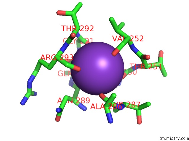
Mono view
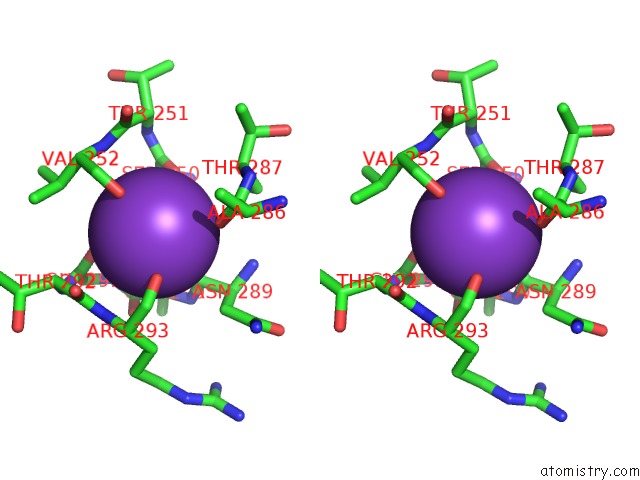
Stereo pair view
Mono view
Stereo pair view
A full contact list of Potassium with other atoms in the K binding
site number 1 of Crystal Structure of the Phosphofructokinase-2 From Escherichia Coli in Complex with Potassium within 5.0Å range:
|
Potassium binding site 2 out of 2 in 3umo
Go back to
Potassium binding site 2 out
of 2 in the Crystal Structure of the Phosphofructokinase-2 From Escherichia Coli in Complex with Potassium
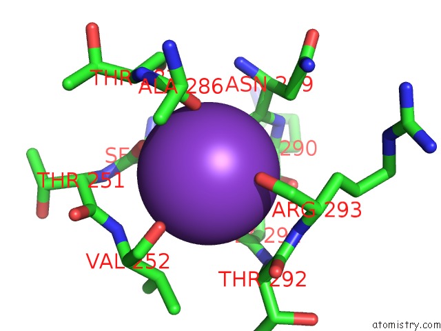
Mono view
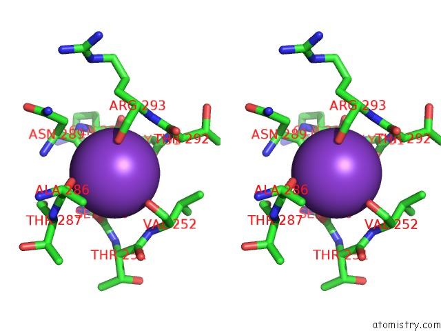
Stereo pair view
Mono view
Stereo pair view
A full contact list of Potassium with other atoms in the K binding
site number 2 of Crystal Structure of the Phosphofructokinase-2 From Escherichia Coli in Complex with Potassium within 5.0Å range:
|
Reference:
M.Baez,
R.Cabrera,
H.M.Pereira,
A.Blanco,
P.Villalobos,
C.A.Ramirez-Sarmiento,
A.Caniuguir,
V.Guixe,
R.C.Garratt,
J.Babul.
A Ribokinase Family Conserved Monovalent Cation Binding Site Enhances the Mgatp-Induced Inhibition in E. Coli Phosphofructokinase-2 Biophys.J. V. 105 185 2013.
ISSN: ISSN 0006-3495
PubMed: 23823238
DOI: 10.1016/J.BPJ.2013.05.028
Page generated: Mon Aug 12 09:43:01 2024
ISSN: ISSN 0006-3495
PubMed: 23823238
DOI: 10.1016/J.BPJ.2013.05.028
Last articles
Fe in 2YXOFe in 2YRS
Fe in 2YXC
Fe in 2YNM
Fe in 2YVJ
Fe in 2YP1
Fe in 2YU2
Fe in 2YU1
Fe in 2YQB
Fe in 2YOO