Potassium »
PDB 3srd-3ugx »
3ss7 »
Potassium in PDB 3ss7: Crystal Structure of Holo D-Serine Dehydratase From Escherichia Coli at 1.55 A Resolution
Enzymatic activity of Crystal Structure of Holo D-Serine Dehydratase From Escherichia Coli at 1.55 A Resolution
All present enzymatic activity of Crystal Structure of Holo D-Serine Dehydratase From Escherichia Coli at 1.55 A Resolution:
4.3.1.18;
4.3.1.18;
Protein crystallography data
The structure of Crystal Structure of Holo D-Serine Dehydratase From Escherichia Coli at 1.55 A Resolution, PDB code: 3ss7
was solved by
D.V.Urusova,
M.N.Isupov,
S.V.Antonyuk,
G.S.Kachalova,
A.A.Vagin,
A.A.Lebedev,
G.P.Bourenkov,
Z.Dauter,
H.D.Bartunik,
W.R.Melik-Adamyan,
T.D.Mueller,
K.D.Schnackerz,
with X-Ray Crystallography technique. A brief refinement statistics is given in the table below:
| Resolution Low / High (Å) | 50.00 / 1.55 |
| Space group | P 1 21 1 |
| Cell size a, b, c (Å), α, β, γ (°) | 73.774, 47.806, 75.193, 90.00, 104.96, 90.00 |
| R / Rfree (%) | 15.9 / 19.4 |
Potassium Binding Sites:
The binding sites of Potassium atom in the Crystal Structure of Holo D-Serine Dehydratase From Escherichia Coli at 1.55 A Resolution
(pdb code 3ss7). This binding sites where shown within
5.0 Angstroms radius around Potassium atom.
In total only one binding site of Potassium was determined in the Crystal Structure of Holo D-Serine Dehydratase From Escherichia Coli at 1.55 A Resolution, PDB code: 3ss7:
In total only one binding site of Potassium was determined in the Crystal Structure of Holo D-Serine Dehydratase From Escherichia Coli at 1.55 A Resolution, PDB code: 3ss7:
Potassium binding site 1 out of 1 in 3ss7
Go back to
Potassium binding site 1 out
of 1 in the Crystal Structure of Holo D-Serine Dehydratase From Escherichia Coli at 1.55 A Resolution
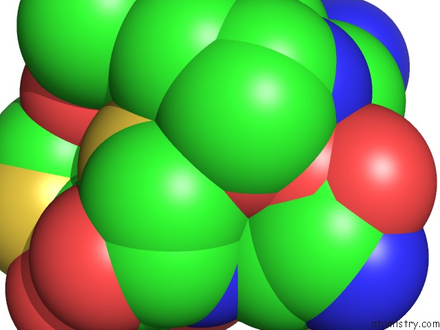
Mono view
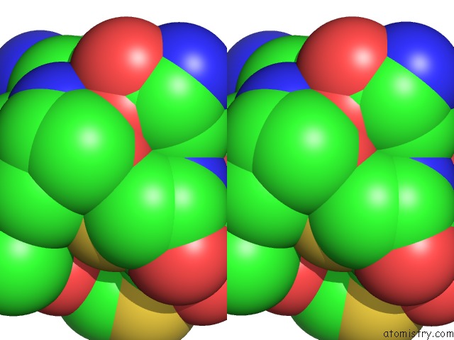
Stereo pair view
Mono view
Stereo pair view
A full contact list of Potassium with other atoms in the K binding
site number 1 of Crystal Structure of Holo D-Serine Dehydratase From Escherichia Coli at 1.55 A Resolution within 5.0Å range:
|
Reference:
D.V.Urusova,
M.N.Isupov,
S.Antonyuk,
G.S.Kachalova,
G.Oblomova,
A.A.Vagin,
A.A.Lebedev,
G.P.Bourenko,
Z.Dauter,
H.D.Bartunik,
V.S.Lamzin,
W.R.Melik-Adamyan,
T.D.Mueller,
K.D.Schnackerz.
Crystal Structure of D-Serine Dehydratase From Escherichia Coli. Biochim.Biophys.Acta V.1824 422 2011.
ISSN: ISSN 0006-3002
PubMed: 22197591
DOI: 10.1016/J.BBAPAP.2011.10.017
Page generated: Sat Aug 9 05:53:23 2025
ISSN: ISSN 0006-3002
PubMed: 22197591
DOI: 10.1016/J.BBAPAP.2011.10.017
Last articles
Mn in 7L3VMn in 7LAF
Mn in 7L3M
Mn in 7L6R
Mn in 7L36
Mn in 7KTM
Mn in 7L29
Mn in 7L28
Mn in 7KTL
Mn in 7L27