Potassium »
PDB 3rs9-3spj »
3s3x »
Potassium in PDB 3s3x: Structure of Chicken Acid-Sensing Ion Channel 1 at 3.0 A Resolution in Complex with Psalmotoxin
Protein crystallography data
The structure of Structure of Chicken Acid-Sensing Ion Channel 1 at 3.0 A Resolution in Complex with Psalmotoxin, PDB code: 3s3x
was solved by
R.J.P.Dawson,
J.Benz,
P.Stohler,
T.Tetaz,
C.Joseph,
S.Huber,
G.Schmid,
D.Huegin,
P.Pflimlin,
G.Trube,
M.G.Rudolph,
M.Hennig,
A.Ruf,
with X-Ray Crystallography technique. A brief refinement statistics is given in the table below:
| Resolution Low / High (Å) | 29.92 / 2.99 |
| Space group | C 1 2 1 |
| Cell size a, b, c (Å), α, β, γ (°) | 232.396, 109.442, 127.269, 90.00, 119.81, 90.00 |
| R / Rfree (%) | 21.8 / 24.9 |
Other elements in 3s3x:
The structure of Structure of Chicken Acid-Sensing Ion Channel 1 at 3.0 A Resolution in Complex with Psalmotoxin also contains other interesting chemical elements:
| Chlorine | (Cl) | 1 atom |
Potassium Binding Sites:
The binding sites of Potassium atom in the Structure of Chicken Acid-Sensing Ion Channel 1 at 3.0 A Resolution in Complex with Psalmotoxin
(pdb code 3s3x). This binding sites where shown within
5.0 Angstroms radius around Potassium atom.
In total 2 binding sites of Potassium where determined in the Structure of Chicken Acid-Sensing Ion Channel 1 at 3.0 A Resolution in Complex with Psalmotoxin, PDB code: 3s3x:
Jump to Potassium binding site number: 1; 2;
In total 2 binding sites of Potassium where determined in the Structure of Chicken Acid-Sensing Ion Channel 1 at 3.0 A Resolution in Complex with Psalmotoxin, PDB code: 3s3x:
Jump to Potassium binding site number: 1; 2;
Potassium binding site 1 out of 2 in 3s3x
Go back to
Potassium binding site 1 out
of 2 in the Structure of Chicken Acid-Sensing Ion Channel 1 at 3.0 A Resolution in Complex with Psalmotoxin
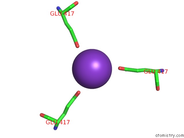
Mono view
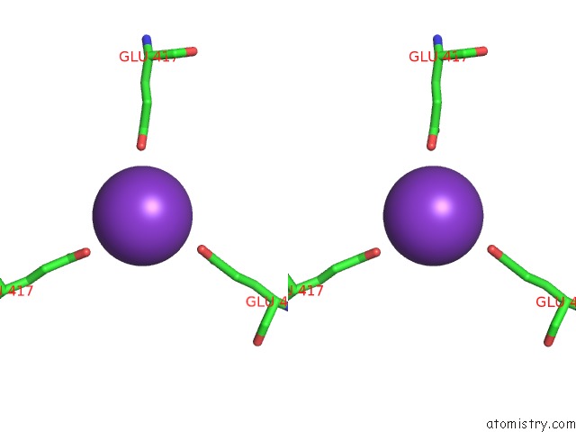
Stereo pair view
Mono view
Stereo pair view
A full contact list of Potassium with other atoms in the K binding
site number 1 of Structure of Chicken Acid-Sensing Ion Channel 1 at 3.0 A Resolution in Complex with Psalmotoxin within 5.0Å range:
|
Potassium binding site 2 out of 2 in 3s3x
Go back to
Potassium binding site 2 out
of 2 in the Structure of Chicken Acid-Sensing Ion Channel 1 at 3.0 A Resolution in Complex with Psalmotoxin
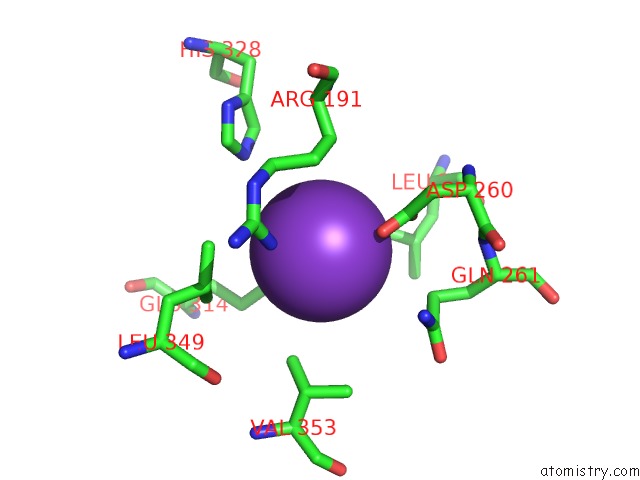
Mono view
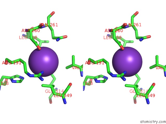
Stereo pair view
Mono view
Stereo pair view
A full contact list of Potassium with other atoms in the K binding
site number 2 of Structure of Chicken Acid-Sensing Ion Channel 1 at 3.0 A Resolution in Complex with Psalmotoxin within 5.0Å range:
|
Reference:
R.J.Dawson,
J.Benz,
P.Stohler,
T.Tetaz,
C.Joseph,
S.Huber,
G.Schmid,
D.Hugin,
P.Pflimlin,
G.Trube,
M.G.Rudolph,
M.Hennig,
A.Ruf.
Structure of the Acid-Sensing Ion Channel 1 in Complex with the Gating Modifier Psalmotoxin 1. Nat Commun V. 3 936 2012.
ISSN: ESSN 2041-1723
PubMed: 22760635
DOI: 10.1038/NCOMMS1917
Page generated: Sat Aug 9 05:46:56 2025
ISSN: ESSN 2041-1723
PubMed: 22760635
DOI: 10.1038/NCOMMS1917
Last articles
Mn in 9LJUMn in 9LJW
Mn in 9LJS
Mn in 9LJR
Mn in 9LJT
Mn in 9LJV
Mg in 9UA2
Mg in 9R96
Mg in 9VM1
Mg in 9P01