Potassium »
PDB 3q9c-3rs8 »
3qlp »
Potassium in PDB 3qlp: X-Ray Structure of the Complex Between Human Alpha Thrombin and A Modified Thrombin Binding Aptamer (Mtba)
Enzymatic activity of X-Ray Structure of the Complex Between Human Alpha Thrombin and A Modified Thrombin Binding Aptamer (Mtba)
All present enzymatic activity of X-Ray Structure of the Complex Between Human Alpha Thrombin and A Modified Thrombin Binding Aptamer (Mtba):
3.4.21.5;
3.4.21.5;
Protein crystallography data
The structure of X-Ray Structure of the Complex Between Human Alpha Thrombin and A Modified Thrombin Binding Aptamer (Mtba), PDB code: 3qlp
was solved by
I.Russo Krauss,
A.Merlino,
L.Mazzarella,
F.Sica,
with X-Ray Crystallography technique. A brief refinement statistics is given in the table below:
| Resolution Low / High (Å) | 25.10 / 2.14 |
| Space group | I 2 2 2 |
| Cell size a, b, c (Å), α, β, γ (°) | 68.208, 110.574, 120.916, 90.00, 90.00, 90.00 |
| R / Rfree (%) | 18.3 / 23.7 |
Other elements in 3qlp:
The structure of X-Ray Structure of the Complex Between Human Alpha Thrombin and A Modified Thrombin Binding Aptamer (Mtba) also contains other interesting chemical elements:
| Sodium | (Na) | 1 atom |
Potassium Binding Sites:
The binding sites of Potassium atom in the X-Ray Structure of the Complex Between Human Alpha Thrombin and A Modified Thrombin Binding Aptamer (Mtba)
(pdb code 3qlp). This binding sites where shown within
5.0 Angstroms radius around Potassium atom.
In total only one binding site of Potassium was determined in the X-Ray Structure of the Complex Between Human Alpha Thrombin and A Modified Thrombin Binding Aptamer (Mtba), PDB code: 3qlp:
In total only one binding site of Potassium was determined in the X-Ray Structure of the Complex Between Human Alpha Thrombin and A Modified Thrombin Binding Aptamer (Mtba), PDB code: 3qlp:
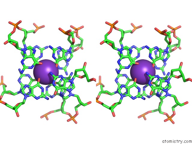
Potassium binding site 1 out of 1 in 3qlp
Go back to
Potassium binding site 1 out
of 1 in the X-Ray Structure of the Complex Between Human Alpha Thrombin and A Modified Thrombin Binding Aptamer (Mtba)
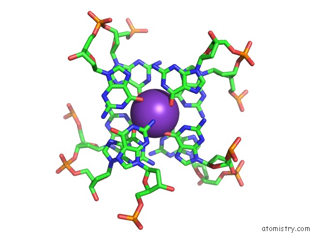
Mono view
Stereo pair view
Mono view
Stereo pair view
A full contact list of Potassium with other atoms in the K binding
site number 1 of X-Ray Structure of the Complex Between Human Alpha Thrombin and A Modified Thrombin Binding Aptamer (Mtba) within 5.0Å range:
|
Reference:
I.Russo Krauss,
A.Merlino,
C.Giancola,
A.Randazzo,
L.Mazzarella,
F.Sica.
Thrombin-Aptamer Recognition: A Revealed Ambiguity. Nucleic Acids Res. V. 39 7858 2011.
ISSN: ISSN 0305-1048
PubMed: 21715374
DOI: 10.1093/NAR/GKR522
Page generated: Sat Aug 9 05:38:38 2025
ISSN: ISSN 0305-1048
PubMed: 21715374
DOI: 10.1093/NAR/GKR522
Last articles
Mn in 2V8TMn in 2VFZ
Mn in 2VBM
Mn in 2V3Z
Mn in 2VBE
Mn in 2V8J
Mn in 2QVW
Mn in 2V3Y
Mn in 2V3X
Mn in 2V09