Potassium »
PDB 3gvx-3irp »
3i6t »
Potassium in PDB 3i6t: Crystal Structure of Muconate Cycloisomerase From Jannaschia Sp.
Protein crystallography data
The structure of Crystal Structure of Muconate Cycloisomerase From Jannaschia Sp., PDB code: 3i6t
was solved by
J.B.Bonanno,
J.Freeman,
K.T.Bain,
J.Do,
R.Romero,
J.M.Sauder,
S.K.Burley,
S.C.Almo,
New York Sgx Research Center For Structural Genomics(Nysgxrc),
with X-Ray Crystallography technique. A brief refinement statistics is given in the table below:
| Resolution Low / High (Å) | 20.00 / 1.90 |
| Space group | P 4 |
| Cell size a, b, c (Å), α, β, γ (°) | 134.196, 134.196, 89.740, 90.00, 90.00, 90.00 |
| R / Rfree (%) | 16.9 / 20.5 |
Other elements in 3i6t:
The structure of Crystal Structure of Muconate Cycloisomerase From Jannaschia Sp. also contains other interesting chemical elements:
| Magnesium | (Mg) | 4 atoms |
Potassium Binding Sites:
The binding sites of Potassium atom in the Crystal Structure of Muconate Cycloisomerase From Jannaschia Sp.
(pdb code 3i6t). This binding sites where shown within
5.0 Angstroms radius around Potassium atom.
In total 4 binding sites of Potassium where determined in the Crystal Structure of Muconate Cycloisomerase From Jannaschia Sp., PDB code: 3i6t:
Jump to Potassium binding site number: 1; 2; 3; 4;
In total 4 binding sites of Potassium where determined in the Crystal Structure of Muconate Cycloisomerase From Jannaschia Sp., PDB code: 3i6t:
Jump to Potassium binding site number: 1; 2; 3; 4;
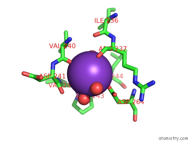
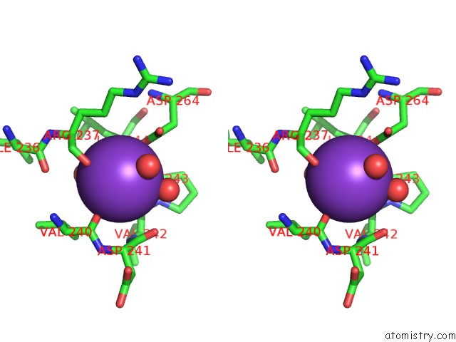
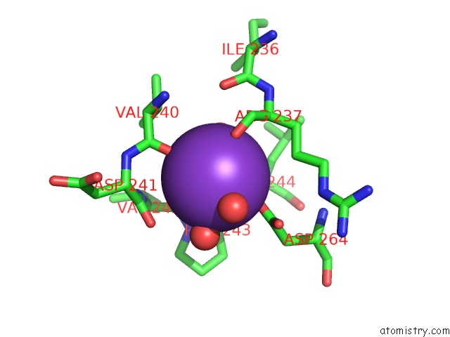
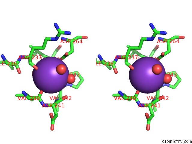
Potassium binding site 1 out of 4 in 3i6t
Go back to
Potassium binding site 1 out
of 4 in the Crystal Structure of Muconate Cycloisomerase From Jannaschia Sp.
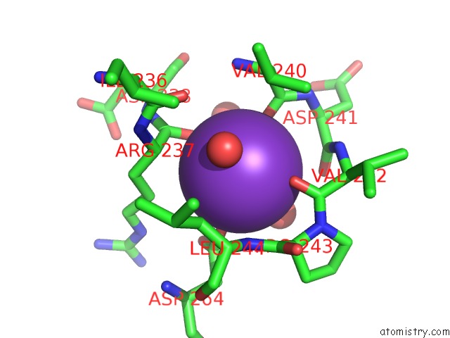
Mono view
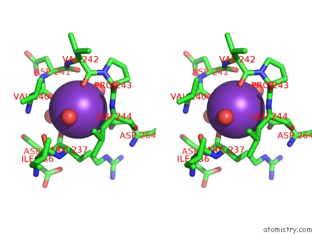
Stereo pair view
Mono view
Stereo pair view
A full contact list of Potassium with other atoms in the K binding
site number 1 of Crystal Structure of Muconate Cycloisomerase From Jannaschia Sp. within 5.0Å range:
|
Potassium binding site 2 out of 4 in 3i6t
Go back to
Potassium binding site 2 out
of 4 in the Crystal Structure of Muconate Cycloisomerase From Jannaschia Sp.
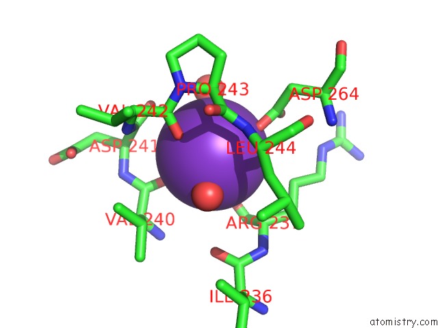
Mono view
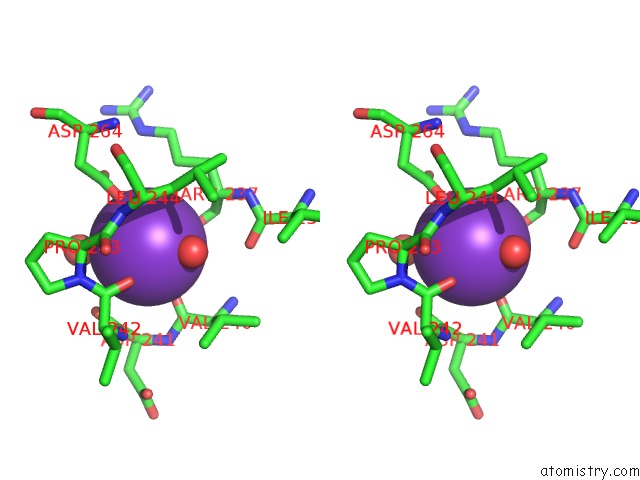
Stereo pair view
Mono view
Stereo pair view
A full contact list of Potassium with other atoms in the K binding
site number 2 of Crystal Structure of Muconate Cycloisomerase From Jannaschia Sp. within 5.0Å range:
|
Potassium binding site 3 out of 4 in 3i6t
Go back to
Potassium binding site 3 out
of 4 in the Crystal Structure of Muconate Cycloisomerase From Jannaschia Sp.
Mono view
Stereo pair view
Mono view
Stereo pair view
A full contact list of Potassium with other atoms in the K binding
site number 3 of Crystal Structure of Muconate Cycloisomerase From Jannaschia Sp. within 5.0Å range:
|
Potassium binding site 4 out of 4 in 3i6t
Go back to
Potassium binding site 4 out
of 4 in the Crystal Structure of Muconate Cycloisomerase From Jannaschia Sp.
Mono view
Stereo pair view
Mono view
Stereo pair view
A full contact list of Potassium with other atoms in the K binding
site number 4 of Crystal Structure of Muconate Cycloisomerase From Jannaschia Sp. within 5.0Å range:
|
Reference:
J.B.Bonanno,
J.Freeman,
K.T.Bain,
J.Do,
R.Romero,
J.M.Sauder,
S.K.Burley,
S.C.Almo.
Crystal Structure of Muconate Cycloisomerase From Jannaschia Sp. To Be Published.
Page generated: Sat Aug 9 05:03:44 2025
Last articles
Mg in 7KXGMg in 7KZ2
Mg in 7KZ4
Mg in 7KYB
Mg in 7KYC
Mg in 7KYA
Mg in 7KXH
Mg in 7KY9
Mg in 7KY8
Mg in 7KY7