Potassium »
PDB 3du0-3f2t »
3exf »
Potassium in PDB 3exf: Crystal Structure of the Pyruvate Dehydrogenase (E1P) Component of Human Pyruvate Dehydrogenase Complex
Enzymatic activity of Crystal Structure of the Pyruvate Dehydrogenase (E1P) Component of Human Pyruvate Dehydrogenase Complex
All present enzymatic activity of Crystal Structure of the Pyruvate Dehydrogenase (E1P) Component of Human Pyruvate Dehydrogenase Complex:
1.2.4.1;
1.2.4.1;
Protein crystallography data
The structure of Crystal Structure of the Pyruvate Dehydrogenase (E1P) Component of Human Pyruvate Dehydrogenase Complex, PDB code: 3exf
was solved by
M.Kato,
R.M.Wynn,
J.L.Chuang,
S.-C.Tso,
M.Machius,
J.Li,
D.T.Chuang,
with X-Ray Crystallography technique. A brief refinement statistics is given in the table below:
| Resolution Low / High (Å) | 50.00 / 3.00 |
| Space group | P 1 21 1 |
| Cell size a, b, c (Å), α, β, γ (°) | 103.389, 129.668, 144.946, 90.00, 109.15, 90.00 |
| R / Rfree (%) | 18.5 / 26.3 |
Other elements in 3exf:
The structure of Crystal Structure of the Pyruvate Dehydrogenase (E1P) Component of Human Pyruvate Dehydrogenase Complex also contains other interesting chemical elements:
| Magnesium | (Mg) | 4 atoms |
Potassium Binding Sites:
The binding sites of Potassium atom in the Crystal Structure of the Pyruvate Dehydrogenase (E1P) Component of Human Pyruvate Dehydrogenase Complex
(pdb code 3exf). This binding sites where shown within
5.0 Angstroms radius around Potassium atom.
In total 4 binding sites of Potassium where determined in the Crystal Structure of the Pyruvate Dehydrogenase (E1P) Component of Human Pyruvate Dehydrogenase Complex, PDB code: 3exf:
Jump to Potassium binding site number: 1; 2; 3; 4;
In total 4 binding sites of Potassium where determined in the Crystal Structure of the Pyruvate Dehydrogenase (E1P) Component of Human Pyruvate Dehydrogenase Complex, PDB code: 3exf:
Jump to Potassium binding site number: 1; 2; 3; 4;
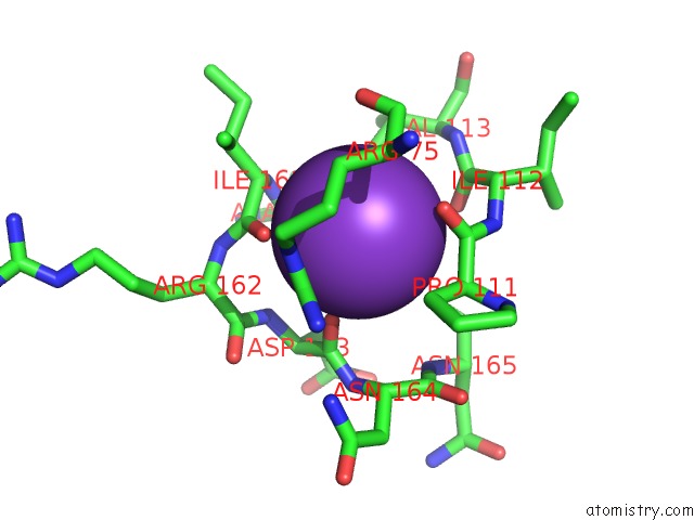
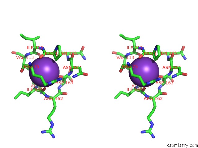
Potassium binding site 1 out of 4 in 3exf
Go back to
Potassium binding site 1 out
of 4 in the Crystal Structure of the Pyruvate Dehydrogenase (E1P) Component of Human Pyruvate Dehydrogenase Complex
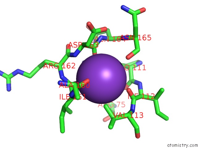
Mono view
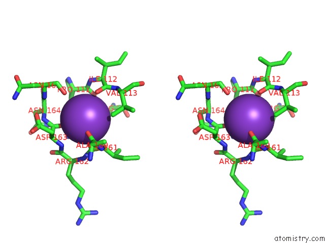
Stereo pair view
Mono view
Stereo pair view
A full contact list of Potassium with other atoms in the K binding
site number 1 of Crystal Structure of the Pyruvate Dehydrogenase (E1P) Component of Human Pyruvate Dehydrogenase Complex within 5.0Å range:
|
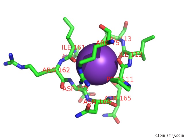
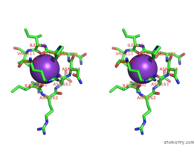
Potassium binding site 2 out of 4 in 3exf
Go back to
Potassium binding site 2 out
of 4 in the Crystal Structure of the Pyruvate Dehydrogenase (E1P) Component of Human Pyruvate Dehydrogenase Complex
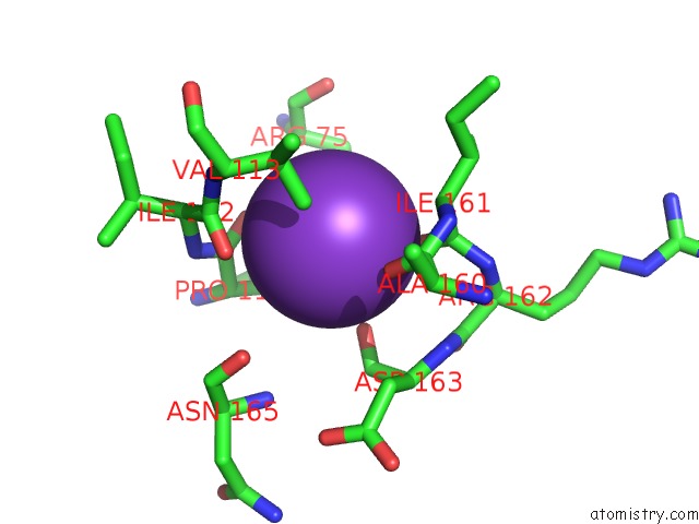
Mono view
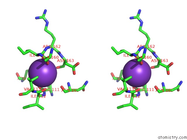
Stereo pair view
Mono view
Stereo pair view
A full contact list of Potassium with other atoms in the K binding
site number 2 of Crystal Structure of the Pyruvate Dehydrogenase (E1P) Component of Human Pyruvate Dehydrogenase Complex within 5.0Å range:
|
Potassium binding site 3 out of 4 in 3exf
Go back to
Potassium binding site 3 out
of 4 in the Crystal Structure of the Pyruvate Dehydrogenase (E1P) Component of Human Pyruvate Dehydrogenase Complex
Mono view
Stereo pair view
Mono view
Stereo pair view
A full contact list of Potassium with other atoms in the K binding
site number 3 of Crystal Structure of the Pyruvate Dehydrogenase (E1P) Component of Human Pyruvate Dehydrogenase Complex within 5.0Å range:
|
Potassium binding site 4 out of 4 in 3exf
Go back to
Potassium binding site 4 out
of 4 in the Crystal Structure of the Pyruvate Dehydrogenase (E1P) Component of Human Pyruvate Dehydrogenase Complex
Mono view
Stereo pair view
Mono view
Stereo pair view
A full contact list of Potassium with other atoms in the K binding
site number 4 of Crystal Structure of the Pyruvate Dehydrogenase (E1P) Component of Human Pyruvate Dehydrogenase Complex within 5.0Å range:
|
Reference:
M.Kato,
R.M.Wynn,
J.L.Chuang,
S.C.Tso,
M.Machius,
J.Li,
D.T.Chuang.
Structural Basis For Inactivation of the Human Pyruvate Dehydrogenase Complex By Phosphorylation: Role of Disordered Phosphorylation Loops. Structure V. 16 1849 2008.
ISSN: ISSN 0969-2126
PubMed: 19081061
DOI: 10.1016/J.STR.2008.10.010
Page generated: Sat Aug 9 04:49:34 2025
ISSN: ISSN 0969-2126
PubMed: 19081061
DOI: 10.1016/J.STR.2008.10.010
Last articles
Na in 6MKBNa in 6ML1
Na in 6MLG
Na in 6MJQ
Na in 6MJI
Na in 6MJA
Na in 6MFM
Na in 6MIY
Na in 6MJ6
Na in 6MIV