Potassium »
PDB 3ccm-3dke »
3den »
Potassium in PDB 3den: Structure of E. Coli Dhdps Mutant Y107W
Enzymatic activity of Structure of E. Coli Dhdps Mutant Y107W
All present enzymatic activity of Structure of E. Coli Dhdps Mutant Y107W:
4.2.1.52;
4.2.1.52;
Protein crystallography data
The structure of Structure of E. Coli Dhdps Mutant Y107W, PDB code: 3den
was solved by
F.G.Pearce,
J.A.Gerrard,
M.A.Perugini,
G.B.Jameson,
with X-Ray Crystallography technique. A brief refinement statistics is given in the table below:
| Resolution Low / High (Å) | 32.22 / 2.20 |
| Space group | P 31 2 1 |
| Cell size a, b, c (Å), α, β, γ (°) | 121.196, 121.196, 110.565, 90.00, 90.00, 120.00 |
| R / Rfree (%) | 18.3 / 24.1 |
Potassium Binding Sites:
The binding sites of Potassium atom in the Structure of E. Coli Dhdps Mutant Y107W
(pdb code 3den). This binding sites where shown within
5.0 Angstroms radius around Potassium atom.
In total 2 binding sites of Potassium where determined in the Structure of E. Coli Dhdps Mutant Y107W, PDB code: 3den:
Jump to Potassium binding site number: 1; 2;
In total 2 binding sites of Potassium where determined in the Structure of E. Coli Dhdps Mutant Y107W, PDB code: 3den:
Jump to Potassium binding site number: 1; 2;
Potassium binding site 1 out of 2 in 3den
Go back to
Potassium binding site 1 out
of 2 in the Structure of E. Coli Dhdps Mutant Y107W
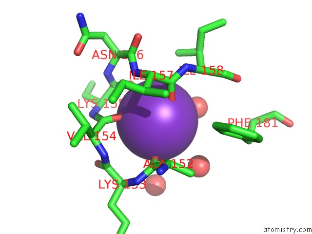
Mono view
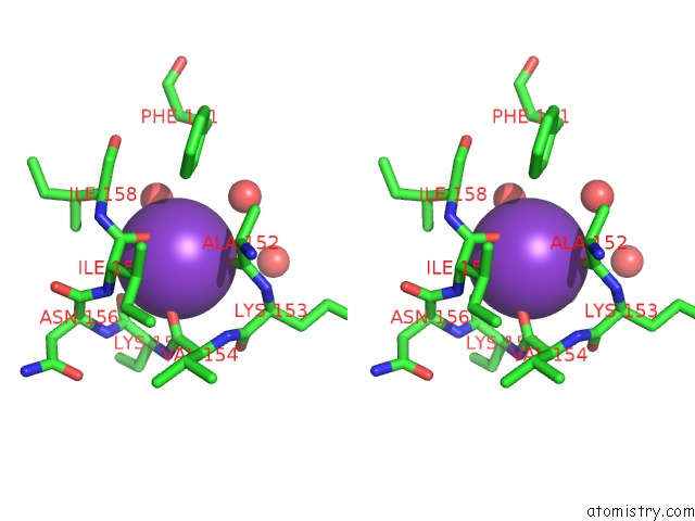
Stereo pair view
Mono view
Stereo pair view
A full contact list of Potassium with other atoms in the K binding
site number 1 of Structure of E. Coli Dhdps Mutant Y107W within 5.0Å range:
|
Potassium binding site 2 out of 2 in 3den
Go back to
Potassium binding site 2 out
of 2 in the Structure of E. Coli Dhdps Mutant Y107W
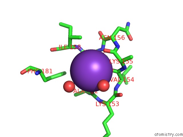
Mono view
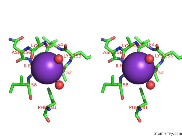
Stereo pair view
Mono view
Stereo pair view
A full contact list of Potassium with other atoms in the K binding
site number 2 of Structure of E. Coli Dhdps Mutant Y107W within 5.0Å range:
|
Reference:
F.G.Pearce,
R.C.J.Dobson,
A.Weber,
L.A.Lane,
M.G.Mccammon,
M.A.Squire,
M.A.Perugini,
G.B.Jameson,
C.V.Robinson,
J.A.Gerrard.
Mutating the Tight-Dimer Interface of Dihydrodipicolinate Synthase Disrupts the Enzyme Quaternary Structure: Toward A Monomeric Enzyme Biochemistry V. 47 12108 2008.
ISSN: ISSN 0006-2960
PubMed: 18937497
DOI: 10.1021/BI801094T
Page generated: Mon Aug 12 08:01:08 2024
ISSN: ISSN 0006-2960
PubMed: 18937497
DOI: 10.1021/BI801094T
Last articles
Fe in 7SH5Fe in 7SHI
Fe in 7SJC
Fe in 7SJB
Fe in 7S3D
Fe in 7SF6
Fe in 7SCP
Fe in 7SBH
Fe in 7S8T
Fe in 7SA3