Potassium »
PDB 2xo1-3atv »
3a45 »
Potassium in PDB 3a45: Crystal Structure of MVNEI1_2
Enzymatic activity of Crystal Structure of MVNEI1_2
All present enzymatic activity of Crystal Structure of MVNEI1_2:
3.2.2.23;
3.2.2.23;
Protein crystallography data
The structure of Crystal Structure of MVNEI1_2, PDB code: 3a45
was solved by
K.Imamura,
S.Wallace,
S.Doublie,
with X-Ray Crystallography technique. A brief refinement statistics is given in the table below:
| Resolution Low / High (Å) | 15.00 / 2.30 |
| Space group | P 1 |
| Cell size a, b, c (Å), α, β, γ (°) | 38.673, 54.417, 87.308, 107.00, 99.86, 93.42 |
| R / Rfree (%) | 20.3 / 26.5 |
Potassium Binding Sites:
The binding sites of Potassium atom in the Crystal Structure of MVNEI1_2
(pdb code 3a45). This binding sites where shown within
5.0 Angstroms radius around Potassium atom.
In total 2 binding sites of Potassium where determined in the Crystal Structure of MVNEI1_2, PDB code: 3a45:
Jump to Potassium binding site number: 1; 2;
In total 2 binding sites of Potassium where determined in the Crystal Structure of MVNEI1_2, PDB code: 3a45:
Jump to Potassium binding site number: 1; 2;
Potassium binding site 1 out of 2 in 3a45
Go back to
Potassium binding site 1 out
of 2 in the Crystal Structure of MVNEI1_2
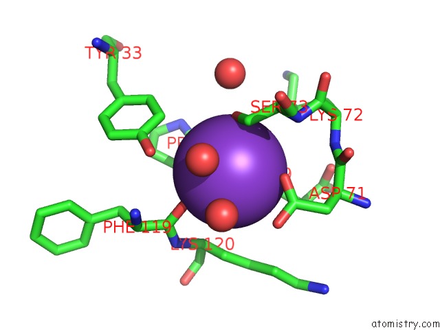
Mono view
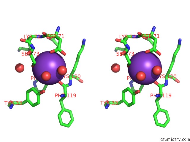
Stereo pair view
Mono view
Stereo pair view
A full contact list of Potassium with other atoms in the K binding
site number 1 of Crystal Structure of MVNEI1_2 within 5.0Å range:
|
Potassium binding site 2 out of 2 in 3a45
Go back to
Potassium binding site 2 out
of 2 in the Crystal Structure of MVNEI1_2
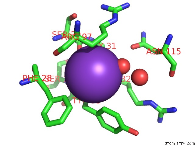
Mono view
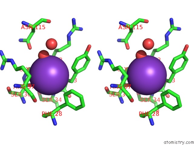
Stereo pair view
Mono view
Stereo pair view
A full contact list of Potassium with other atoms in the K binding
site number 2 of Crystal Structure of MVNEI1_2 within 5.0Å range:
|
Reference:
K.Imamura,
S.S.Wallace,
S.Doublie.
Structural Characterization of A Viral NEIL1 Ortholog Unliganded and Bound to Abasic Site-Containing Dna J.Biol.Chem. V. 284 26174 2009.
ISSN: ISSN 0021-9258
PubMed: 19625256
DOI: 10.1074/JBC.M109.021907
Page generated: Sat Aug 9 04:30:49 2025
ISSN: ISSN 0021-9258
PubMed: 19625256
DOI: 10.1074/JBC.M109.021907
Last articles
Mn in 4QKDMn in 4QKB
Mn in 4QNJ
Mn in 4QKF
Mn in 4QKN
Mn in 4Q42
Mn in 4Q41
Mn in 4QAX
Mn in 4QH9
Mn in 4QAG