Potassium »
PDB 2w0f-2xo0 »
2whv »
Potassium in PDB 2whv: Crystal Structure of Mouse Cadherin-23 EC1-2 (All Cation Binding Sites Occupied By Calcium)
Protein crystallography data
The structure of Crystal Structure of Mouse Cadherin-23 EC1-2 (All Cation Binding Sites Occupied By Calcium), PDB code: 2whv
was solved by
M.Sotomayor,
W.Weihofen,
R.Gaudet,
D.P.Corey,
with X-Ray Crystallography technique. A brief refinement statistics is given in the table below:
| Resolution Low / High (Å) | 22.25 / 2.36 |
| Space group | H 3 2 |
| Cell size a, b, c (Å), α, β, γ (°) | 151.290, 151.290, 136.881, 90.00, 90.00, 120.00 |
| R / Rfree (%) | 20 / 22.1 |
Other elements in 2whv:
The structure of Crystal Structure of Mouse Cadherin-23 EC1-2 (All Cation Binding Sites Occupied By Calcium) also contains other interesting chemical elements:
| Chlorine | (Cl) | 1 atom |
| Calcium | (Ca) | 4 atoms |
Potassium Binding Sites:
The binding sites of Potassium atom in the Crystal Structure of Mouse Cadherin-23 EC1-2 (All Cation Binding Sites Occupied By Calcium)
(pdb code 2whv). This binding sites where shown within
5.0 Angstroms radius around Potassium atom.
In total 4 binding sites of Potassium where determined in the Crystal Structure of Mouse Cadherin-23 EC1-2 (All Cation Binding Sites Occupied By Calcium), PDB code: 2whv:
Jump to Potassium binding site number: 1; 2; 3; 4;
In total 4 binding sites of Potassium where determined in the Crystal Structure of Mouse Cadherin-23 EC1-2 (All Cation Binding Sites Occupied By Calcium), PDB code: 2whv:
Jump to Potassium binding site number: 1; 2; 3; 4;
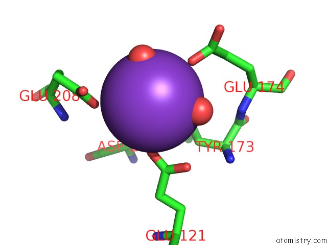
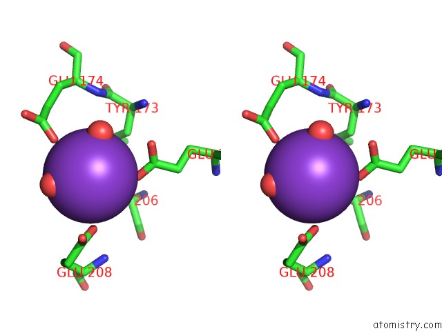
Potassium binding site 1 out of 4 in 2whv
Go back to
Potassium binding site 1 out
of 4 in the Crystal Structure of Mouse Cadherin-23 EC1-2 (All Cation Binding Sites Occupied By Calcium)
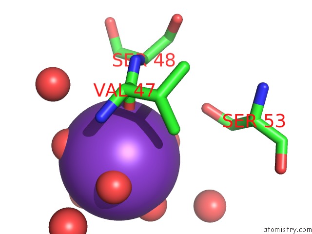
Mono view
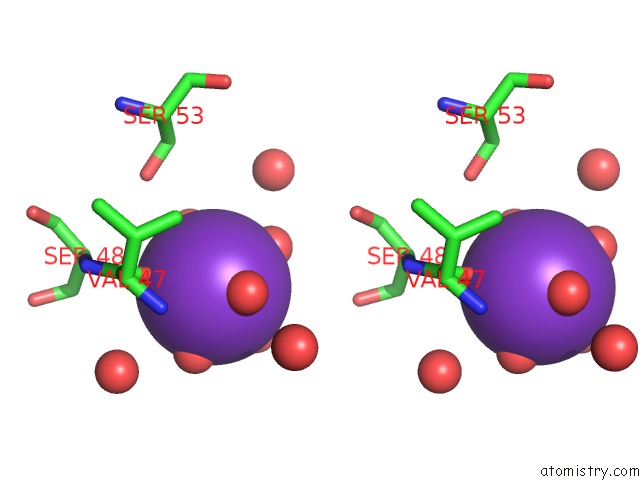
Stereo pair view
Mono view
Stereo pair view
A full contact list of Potassium with other atoms in the K binding
site number 1 of Crystal Structure of Mouse Cadherin-23 EC1-2 (All Cation Binding Sites Occupied By Calcium) within 5.0Å range:
|
Potassium binding site 2 out of 4 in 2whv
Go back to
Potassium binding site 2 out
of 4 in the Crystal Structure of Mouse Cadherin-23 EC1-2 (All Cation Binding Sites Occupied By Calcium)
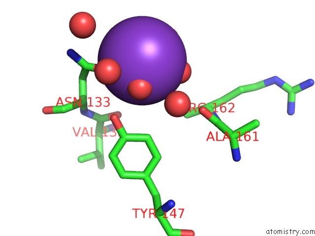
Mono view
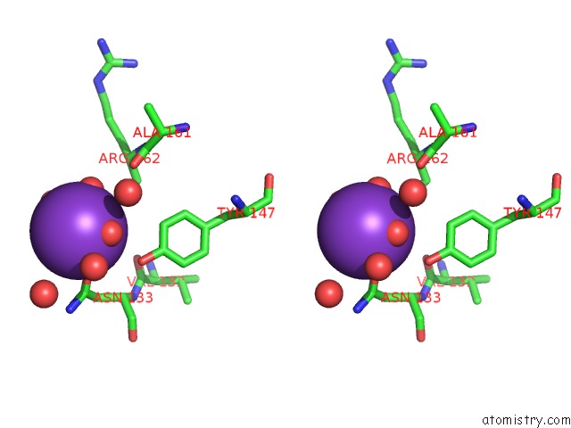
Stereo pair view
Mono view
Stereo pair view
A full contact list of Potassium with other atoms in the K binding
site number 2 of Crystal Structure of Mouse Cadherin-23 EC1-2 (All Cation Binding Sites Occupied By Calcium) within 5.0Å range:
|
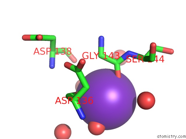
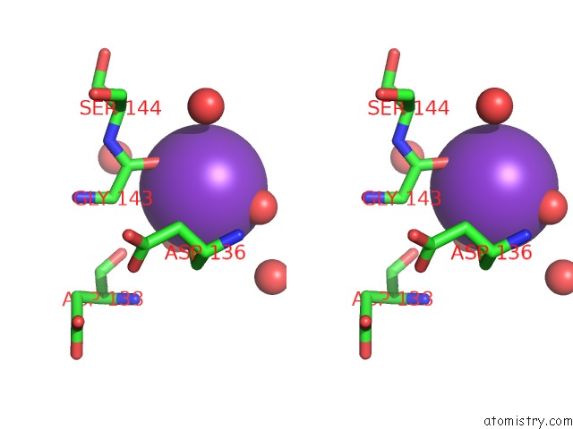
Potassium binding site 3 out of 4 in 2whv
Go back to
Potassium binding site 3 out
of 4 in the Crystal Structure of Mouse Cadherin-23 EC1-2 (All Cation Binding Sites Occupied By Calcium)
Mono view
Stereo pair view
Mono view
Stereo pair view
A full contact list of Potassium with other atoms in the K binding
site number 3 of Crystal Structure of Mouse Cadherin-23 EC1-2 (All Cation Binding Sites Occupied By Calcium) within 5.0Å range:
|
Potassium binding site 4 out of 4 in 2whv
Go back to
Potassium binding site 4 out
of 4 in the Crystal Structure of Mouse Cadherin-23 EC1-2 (All Cation Binding Sites Occupied By Calcium)
Mono view
Stereo pair view
Mono view
Stereo pair view
A full contact list of Potassium with other atoms in the K binding
site number 4 of Crystal Structure of Mouse Cadherin-23 EC1-2 (All Cation Binding Sites Occupied By Calcium) within 5.0Å range:
|
Reference:
M.Sotomayor,
W.Weihofen,
R.Gaudet,
D.P.Corey.
Structural Determinants of Cadherin-23 Function in Hearing and Deafness. Neuron V. 66 85 2010.
ISSN: ISSN 0896-6273
PubMed: 20399731
DOI: 10.1016/J.NEURON.2010.03.028
Page generated: Mon Aug 12 07:28:43 2024
ISSN: ISSN 0896-6273
PubMed: 20399731
DOI: 10.1016/J.NEURON.2010.03.028
Last articles
I in 4S2VI in 4S2M
I in 4TKI
I in 4TQD
I in 4TMD
I in 4S3Q
I in 4TJV
I in 4S22
I in 4S2H
I in 4QX5