Potassium »
PDB 2o8l-2qxl »
2qks »
Potassium in PDB 2qks: Crystal Structure of A KIR3.1-Prokaryotic Kir Channel Chimera
Protein crystallography data
The structure of Crystal Structure of A KIR3.1-Prokaryotic Kir Channel Chimera, PDB code: 2qks
was solved by
M.Nishida,
R.Mackinnon,
with X-Ray Crystallography technique. A brief refinement statistics is given in the table below:
| Resolution Low / High (Å) | 29.50 / 2.20 |
| Space group | P 4 |
| Cell size a, b, c (Å), α, β, γ (°) | 98.416, 98.416, 92.624, 90.00, 90.00, 90.00 |
| R / Rfree (%) | 23.1 / 25.6 |
Potassium Binding Sites:
Pages:
>>> Page 1 <<< Page 2, Binding sites: 11 - 12;Binding sites:
The binding sites of Potassium atom in the Crystal Structure of A KIR3.1-Prokaryotic Kir Channel Chimera (pdb code 2qks). This binding sites where shown within 5.0 Angstroms radius around Potassium atom.In total 12 binding sites of Potassium where determined in the Crystal Structure of A KIR3.1-Prokaryotic Kir Channel Chimera, PDB code: 2qks:
Jump to Potassium binding site number: 1; 2; 3; 4; 5; 6; 7; 8; 9; 10;
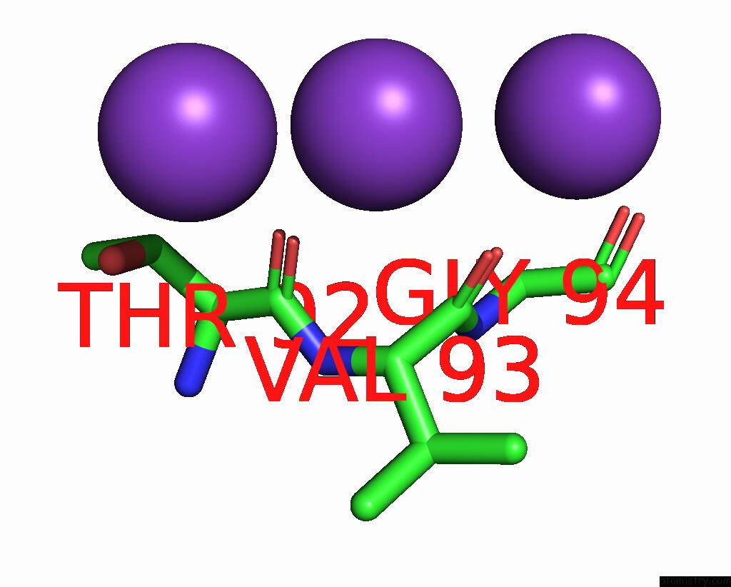
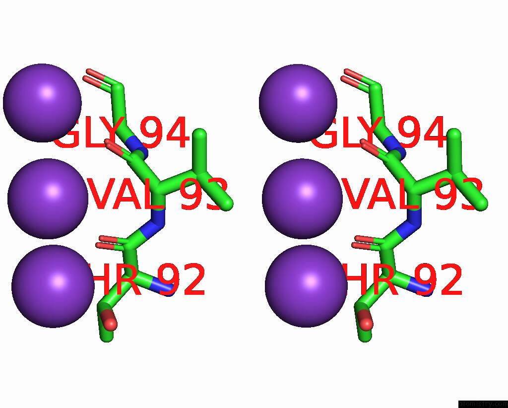
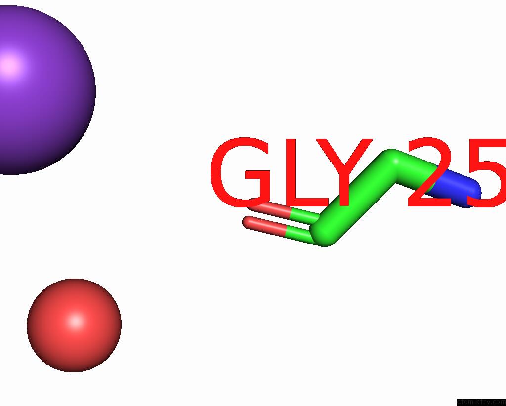
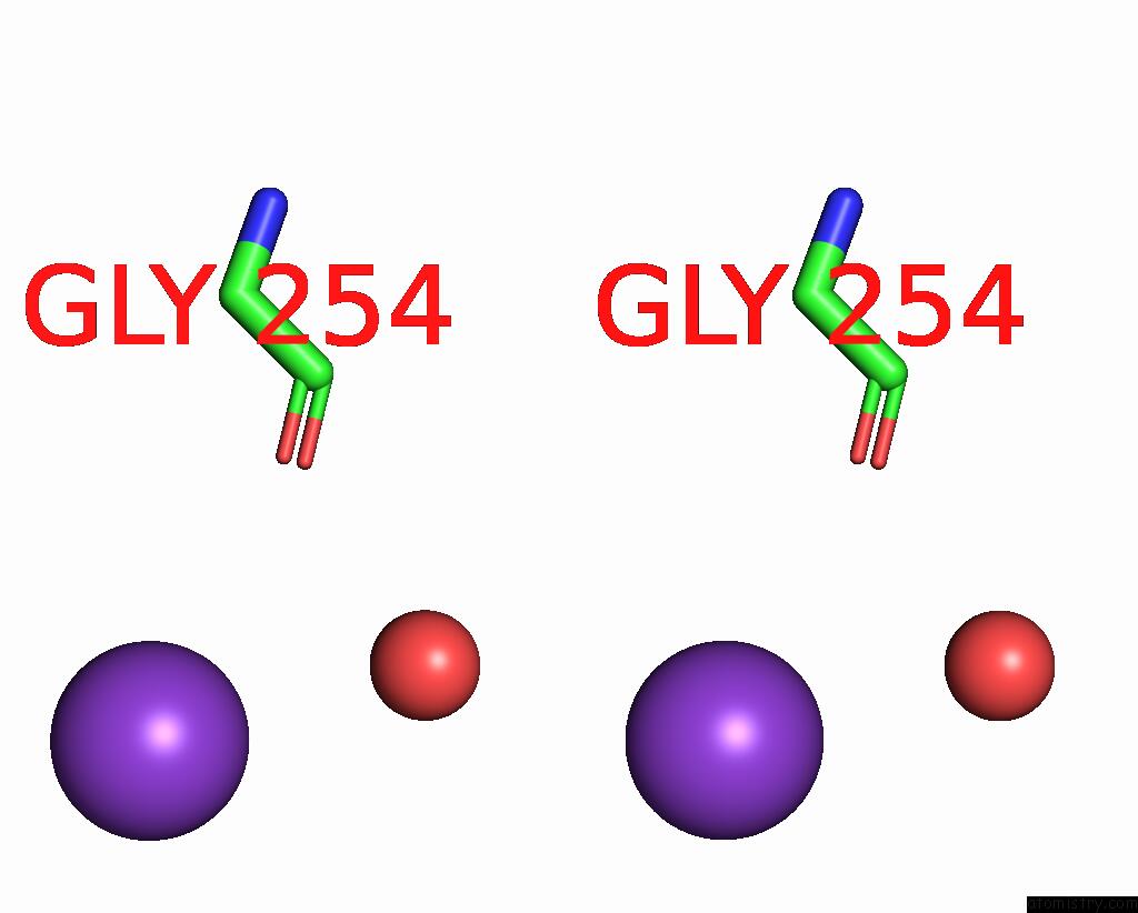
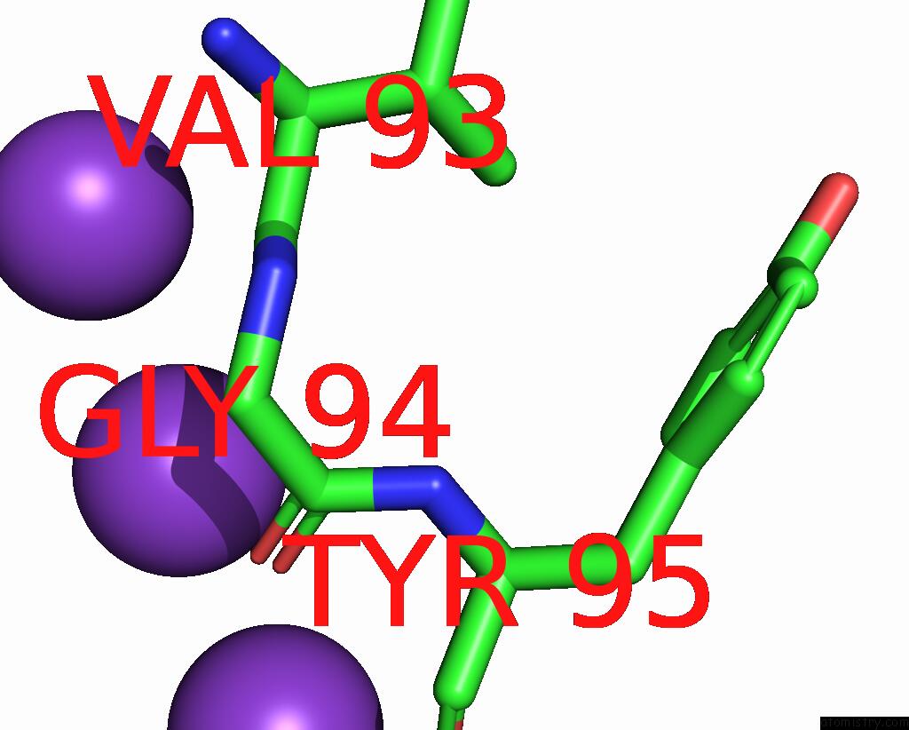
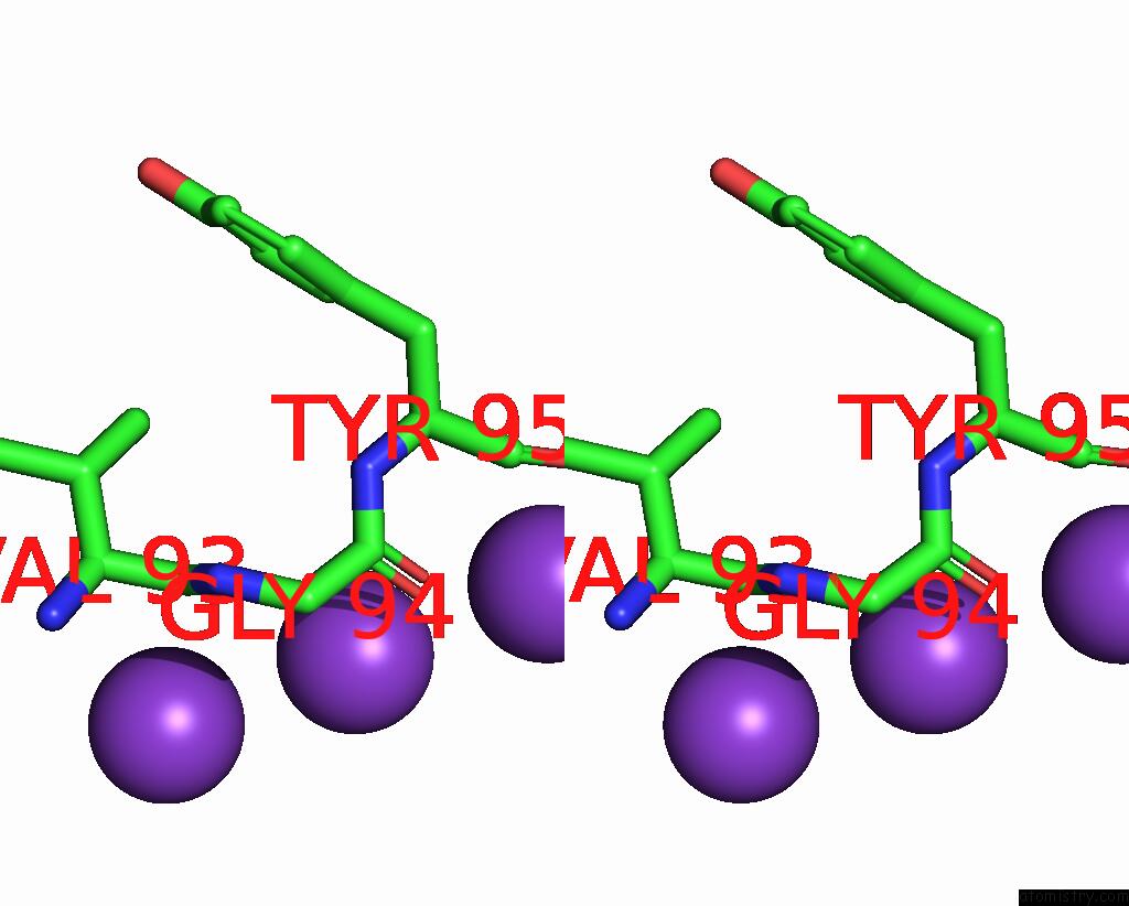
Potassium binding site 1 out of 12 in 2qks
Go back to
Potassium binding site 1 out
of 12 in the Crystal Structure of A KIR3.1-Prokaryotic Kir Channel Chimera
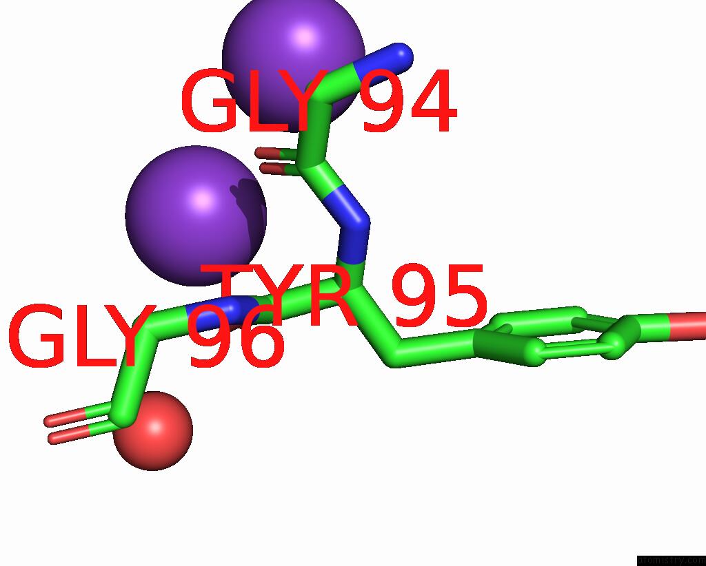
Mono view
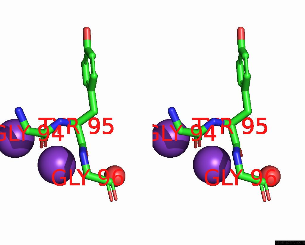
Stereo pair view
Mono view
Stereo pair view
A full contact list of Potassium with other atoms in the K binding
site number 1 of Crystal Structure of A KIR3.1-Prokaryotic Kir Channel Chimera within 5.0Å range:
|
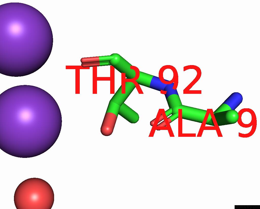
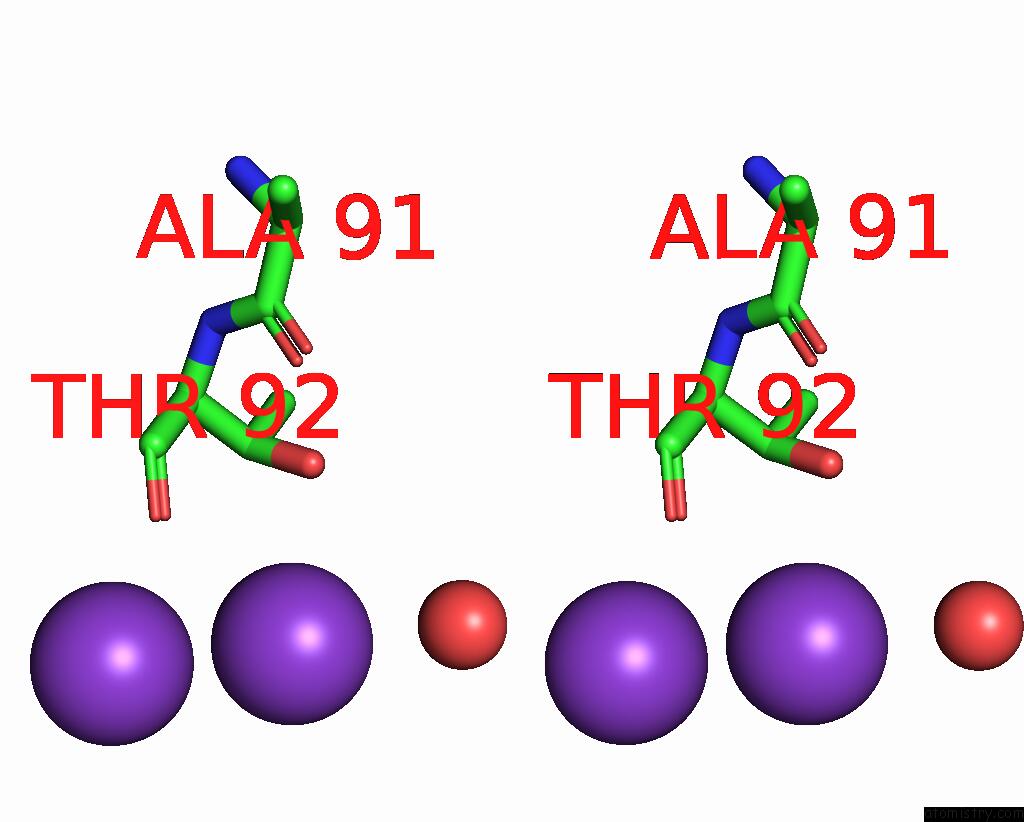
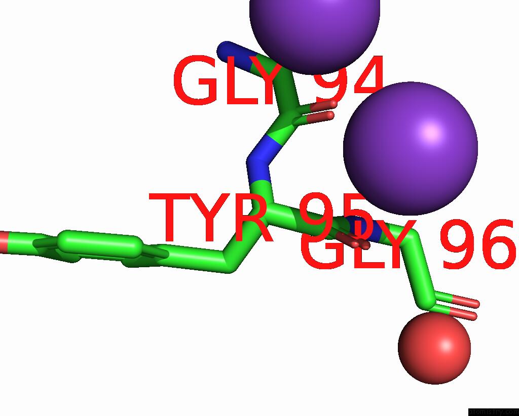
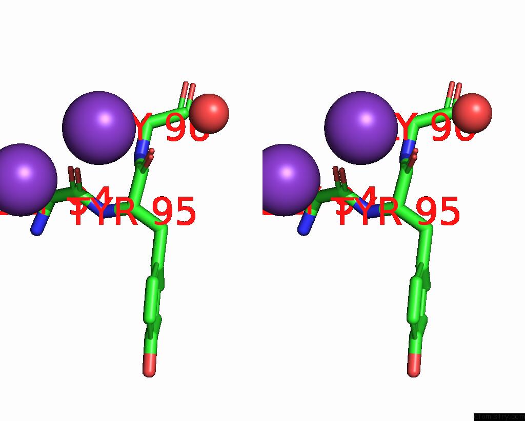
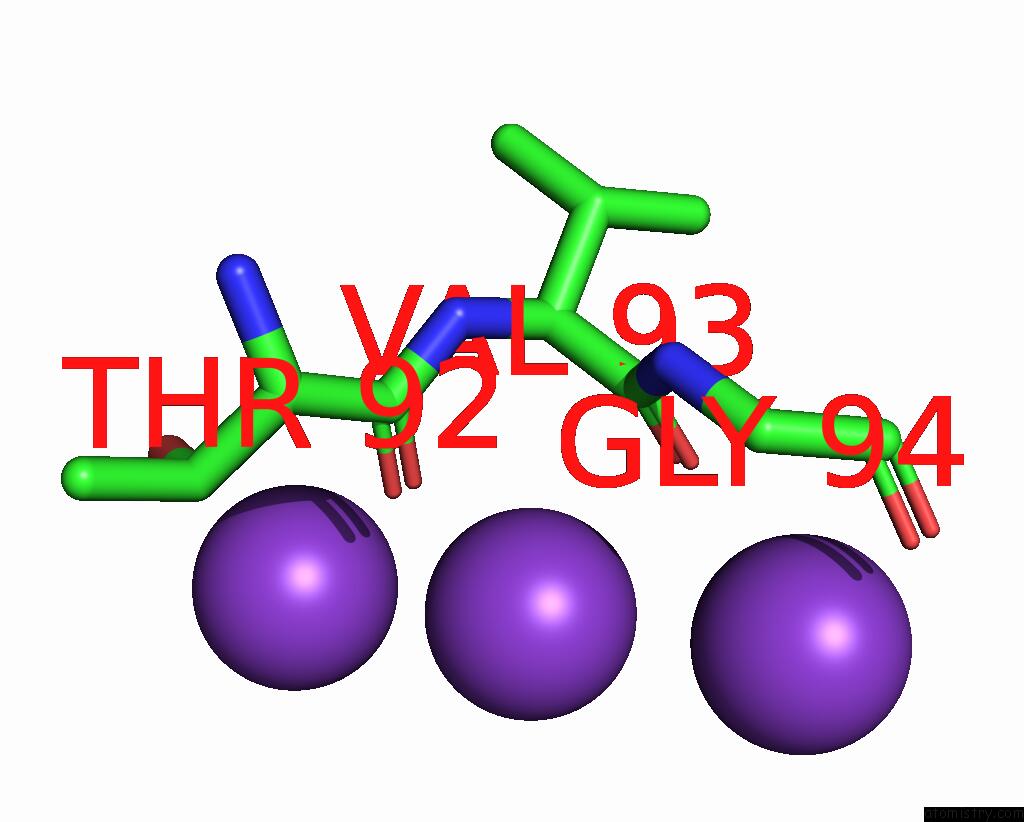
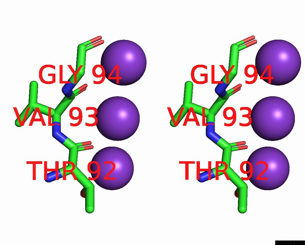
Potassium binding site 2 out of 12 in 2qks
Go back to
Potassium binding site 2 out
of 12 in the Crystal Structure of A KIR3.1-Prokaryotic Kir Channel Chimera
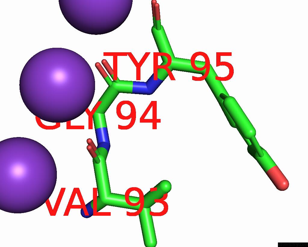
Mono view
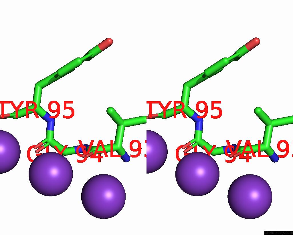
Stereo pair view
Mono view
Stereo pair view
A full contact list of Potassium with other atoms in the K binding
site number 2 of Crystal Structure of A KIR3.1-Prokaryotic Kir Channel Chimera within 5.0Å range:
|
Potassium binding site 3 out of 12 in 2qks
Go back to
Potassium binding site 3 out
of 12 in the Crystal Structure of A KIR3.1-Prokaryotic Kir Channel Chimera
Mono view
Stereo pair view
Mono view
Stereo pair view
A full contact list of Potassium with other atoms in the K binding
site number 3 of Crystal Structure of A KIR3.1-Prokaryotic Kir Channel Chimera within 5.0Å range:
|
Potassium binding site 4 out of 12 in 2qks
Go back to
Potassium binding site 4 out
of 12 in the Crystal Structure of A KIR3.1-Prokaryotic Kir Channel Chimera
Mono view
Stereo pair view
Mono view
Stereo pair view
A full contact list of Potassium with other atoms in the K binding
site number 4 of Crystal Structure of A KIR3.1-Prokaryotic Kir Channel Chimera within 5.0Å range:
|
Potassium binding site 5 out of 12 in 2qks
Go back to
Potassium binding site 5 out
of 12 in the Crystal Structure of A KIR3.1-Prokaryotic Kir Channel Chimera
Mono view
Stereo pair view
Mono view
Stereo pair view
A full contact list of Potassium with other atoms in the K binding
site number 5 of Crystal Structure of A KIR3.1-Prokaryotic Kir Channel Chimera within 5.0Å range:
|
Potassium binding site 6 out of 12 in 2qks
Go back to
Potassium binding site 6 out
of 12 in the Crystal Structure of A KIR3.1-Prokaryotic Kir Channel Chimera
Mono view
Stereo pair view
Mono view
Stereo pair view
A full contact list of Potassium with other atoms in the K binding
site number 6 of Crystal Structure of A KIR3.1-Prokaryotic Kir Channel Chimera within 5.0Å range:
|
Potassium binding site 7 out of 12 in 2qks
Go back to
Potassium binding site 7 out
of 12 in the Crystal Structure of A KIR3.1-Prokaryotic Kir Channel Chimera
Mono view
Stereo pair view
Mono view
Stereo pair view
A full contact list of Potassium with other atoms in the K binding
site number 7 of Crystal Structure of A KIR3.1-Prokaryotic Kir Channel Chimera within 5.0Å range:
|
Potassium binding site 8 out of 12 in 2qks
Go back to
Potassium binding site 8 out
of 12 in the Crystal Structure of A KIR3.1-Prokaryotic Kir Channel Chimera
Mono view
Stereo pair view
Mono view
Stereo pair view
A full contact list of Potassium with other atoms in the K binding
site number 8 of Crystal Structure of A KIR3.1-Prokaryotic Kir Channel Chimera within 5.0Å range:
|
Potassium binding site 9 out of 12 in 2qks
Go back to
Potassium binding site 9 out
of 12 in the Crystal Structure of A KIR3.1-Prokaryotic Kir Channel Chimera
Mono view
Stereo pair view
Mono view
Stereo pair view
A full contact list of Potassium with other atoms in the K binding
site number 9 of Crystal Structure of A KIR3.1-Prokaryotic Kir Channel Chimera within 5.0Å range:
|
Potassium binding site 10 out of 12 in 2qks
Go back to
Potassium binding site 10 out
of 12 in the Crystal Structure of A KIR3.1-Prokaryotic Kir Channel Chimera
Mono view
Stereo pair view
Mono view
Stereo pair view
A full contact list of Potassium with other atoms in the K binding
site number 10 of Crystal Structure of A KIR3.1-Prokaryotic Kir Channel Chimera within 5.0Å range:
|
Reference:
M.Nishida,
M.Cadene,
B.T.Chait,
R.Mackinnon.
Crystal Structure of A KIR3.1-Prokaryotic Kir Channel Chimera. Embo J. V. 26 4005 2007.
ISSN: ISSN 0261-4189
PubMed: 17703190
DOI: 10.1038/SJ.EMBOJ.7601828
Page generated: Sat Aug 9 03:49:44 2025
ISSN: ISSN 0261-4189
PubMed: 17703190
DOI: 10.1038/SJ.EMBOJ.7601828
Last articles
Mn in 5WFWMn in 5WFM
Mn in 5WF3
Mn in 5WEY
Mn in 5WEI
Mn in 5WEB
Mn in 5WEF
Mn in 5WDY
Mn in 5WE8
Mn in 5WE7