Potassium »
PDB 2o8l-2qxl »
2qbn »
Potassium in PDB 2qbn: Crystal Structure of Ferric G248V Cytochrome P450CAM
Enzymatic activity of Crystal Structure of Ferric G248V Cytochrome P450CAM
All present enzymatic activity of Crystal Structure of Ferric G248V Cytochrome P450CAM:
1.14.15.1;
1.14.15.1;
Protein crystallography data
The structure of Crystal Structure of Ferric G248V Cytochrome P450CAM, PDB code: 2qbn
was solved by
K.Von Koenig,
T.M.Makris,
S.D.Sligar,
I.Schlichting,
with X-Ray Crystallography technique. A brief refinement statistics is given in the table below:
| Resolution Low / High (Å) | 20.00 / 1.75 |
| Space group | P 43 21 2 |
| Cell size a, b, c (Å), α, β, γ (°) | 63.920, 63.920, 242.140, 90.00, 90.00, 90.00 |
| R / Rfree (%) | 19.6 / 22.3 |
Other elements in 2qbn:
The structure of Crystal Structure of Ferric G248V Cytochrome P450CAM also contains other interesting chemical elements:
| Iron | (Fe) | 1 atom |
Potassium Binding Sites:
The binding sites of Potassium atom in the Crystal Structure of Ferric G248V Cytochrome P450CAM
(pdb code 2qbn). This binding sites where shown within
5.0 Angstroms radius around Potassium atom.
In total only one binding site of Potassium was determined in the Crystal Structure of Ferric G248V Cytochrome P450CAM, PDB code: 2qbn:
In total only one binding site of Potassium was determined in the Crystal Structure of Ferric G248V Cytochrome P450CAM, PDB code: 2qbn:
Potassium binding site 1 out of 1 in 2qbn
Go back to
Potassium binding site 1 out
of 1 in the Crystal Structure of Ferric G248V Cytochrome P450CAM
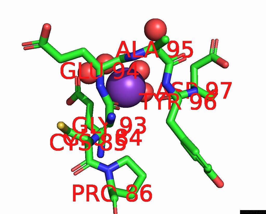
Mono view
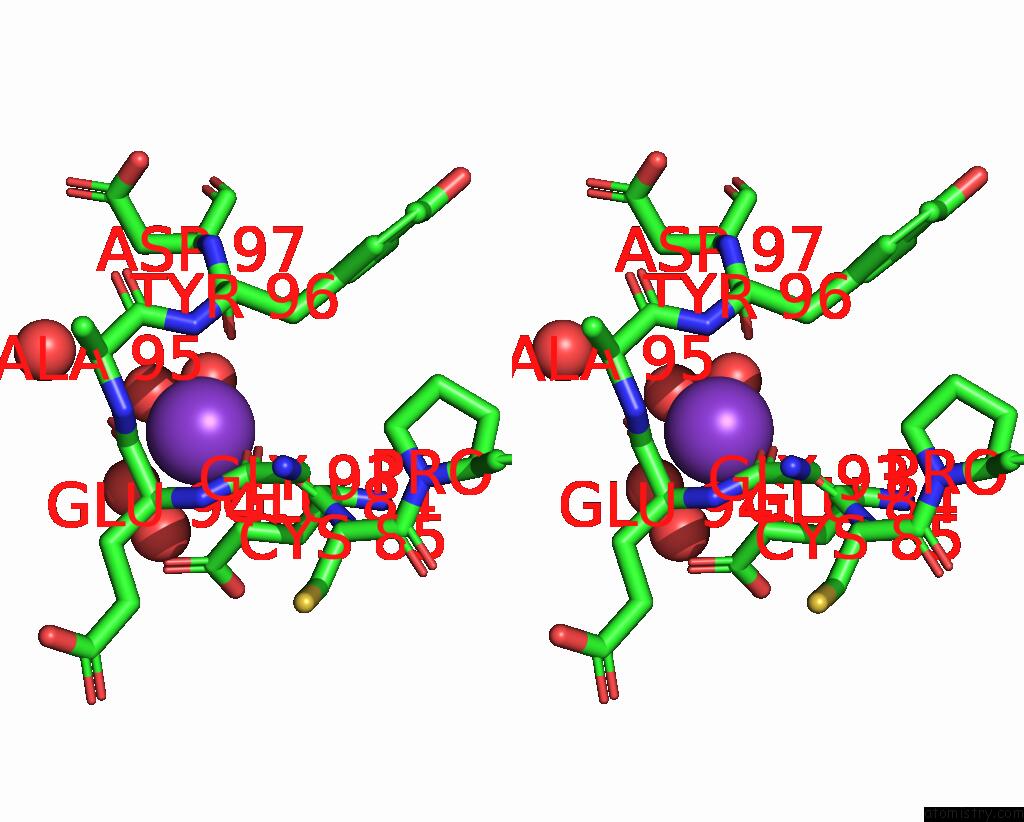
Stereo pair view
Mono view
Stereo pair view
A full contact list of Potassium with other atoms in the K binding
site number 1 of Crystal Structure of Ferric G248V Cytochrome P450CAM within 5.0Å range:
|
Reference:
T.M.Makris,
K.V.Koenig,
I.Schlichting,
S.G.Sligar.
Alteration of P450 Distal Pocket Solvent Leads to Impaired Proton Delivery and Changes in Heme Geometry. Biochemistry V. 46 14129 2007.
ISSN: ISSN 0006-2960
PubMed: 18001135
DOI: 10.1021/BI7013695
Page generated: Sat Aug 9 03:48:33 2025
ISSN: ISSN 0006-2960
PubMed: 18001135
DOI: 10.1021/BI7013695
Last articles
Mg in 3CC2Mg in 3CCE
Mg in 3CC7
Mg in 3CC4
Mg in 3CB3
Mg in 3CC6
Mg in 3C9U
Mg in 3CBT
Mg in 3CBQ
Mg in 3CBG