Potassium »
PDB 2o8l-2qxl »
2q01 »
Potassium in PDB 2q01: Crystal Structure of Glucuronate Isomerase From Caulobacter Crescentus
Enzymatic activity of Crystal Structure of Glucuronate Isomerase From Caulobacter Crescentus
All present enzymatic activity of Crystal Structure of Glucuronate Isomerase From Caulobacter Crescentus:
5.3.1.12;
5.3.1.12;
Protein crystallography data
The structure of Crystal Structure of Glucuronate Isomerase From Caulobacter Crescentus, PDB code: 2q01
was solved by
Y.Patskovsky,
J.Bonanno,
V.Sridhar,
J.M.Sauder,
J.Freeman,
A.Powell,
J.Koss,
C.Groshong,
T.Gheyi,
S.R.Wasserman,
F.Raushel,
S.K.Burley,
S.C.Almo,
New York Sgx Research Center For Structural Genomics(Nysgxrc),
with X-Ray Crystallography technique. A brief refinement statistics is given in the table below:
| Resolution Low / High (Å) | 20.00 / 2.34 |
| Space group | F 2 2 2 |
| Cell size a, b, c (Å), α, β, γ (°) | 177.332, 190.176, 319.290, 90.00, 90.00, 90.00 |
| R / Rfree (%) | 19.8 / 25.1 |
Potassium Binding Sites:
The binding sites of Potassium atom in the Crystal Structure of Glucuronate Isomerase From Caulobacter Crescentus
(pdb code 2q01). This binding sites where shown within
5.0 Angstroms radius around Potassium atom.
In total 3 binding sites of Potassium where determined in the Crystal Structure of Glucuronate Isomerase From Caulobacter Crescentus, PDB code: 2q01:
Jump to Potassium binding site number: 1; 2; 3;
In total 3 binding sites of Potassium where determined in the Crystal Structure of Glucuronate Isomerase From Caulobacter Crescentus, PDB code: 2q01:
Jump to Potassium binding site number: 1; 2; 3;
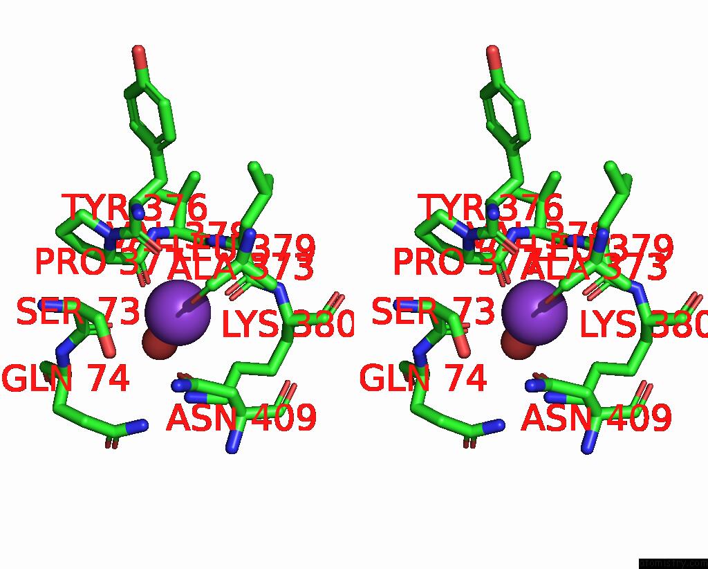
Potassium binding site 1 out of 3 in 2q01
Go back to
Potassium binding site 1 out
of 3 in the Crystal Structure of Glucuronate Isomerase From Caulobacter Crescentus
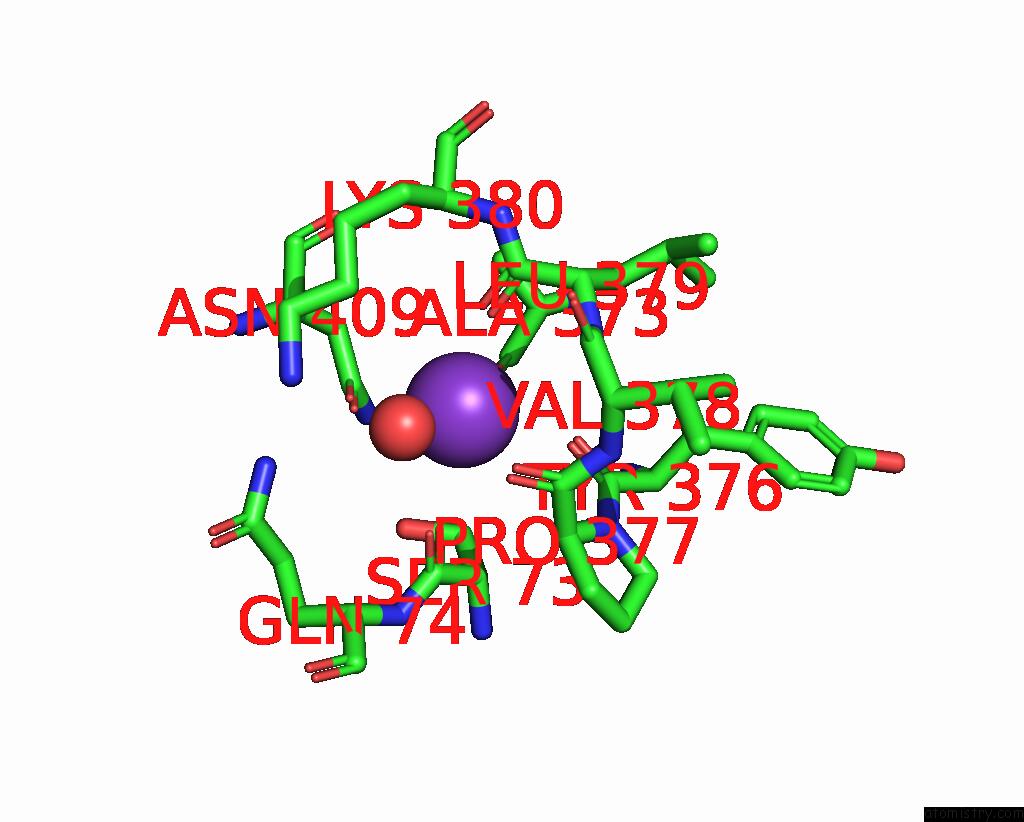
Mono view
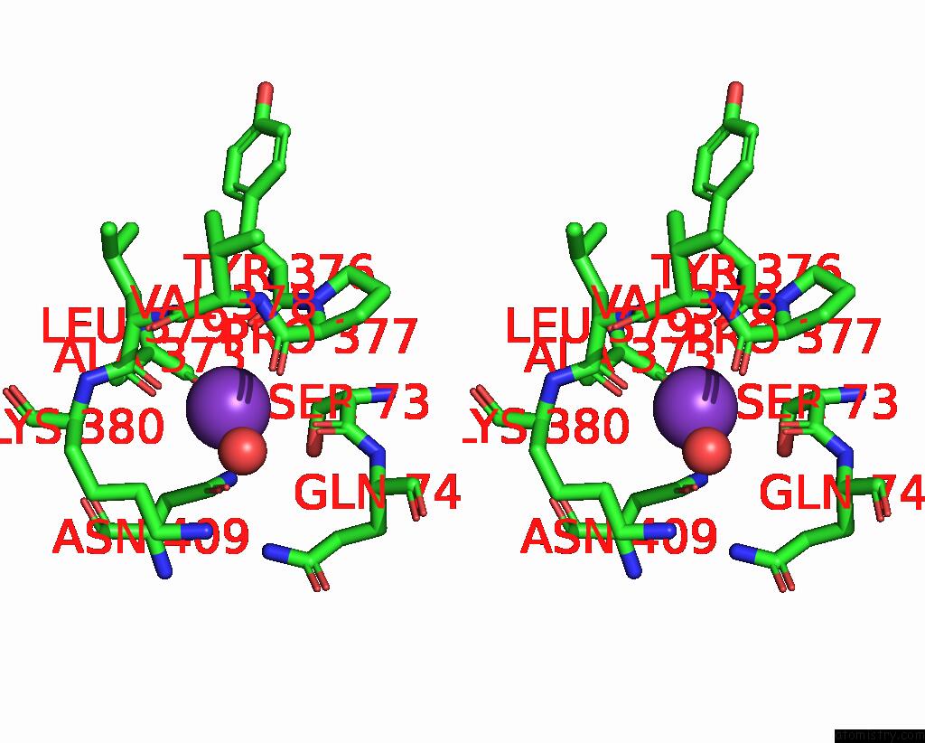
Stereo pair view
Mono view
Stereo pair view
A full contact list of Potassium with other atoms in the K binding
site number 1 of Crystal Structure of Glucuronate Isomerase From Caulobacter Crescentus within 5.0Å range:
|
Potassium binding site 2 out of 3 in 2q01
Go back to
Potassium binding site 2 out
of 3 in the Crystal Structure of Glucuronate Isomerase From Caulobacter Crescentus
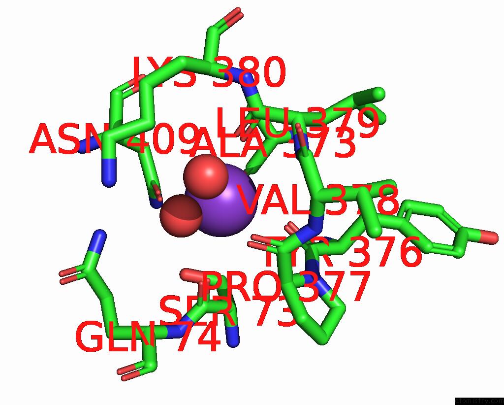
Mono view

Stereo pair view
Mono view
Stereo pair view
A full contact list of Potassium with other atoms in the K binding
site number 2 of Crystal Structure of Glucuronate Isomerase From Caulobacter Crescentus within 5.0Å range:
|
Potassium binding site 3 out of 3 in 2q01
Go back to
Potassium binding site 3 out
of 3 in the Crystal Structure of Glucuronate Isomerase From Caulobacter Crescentus
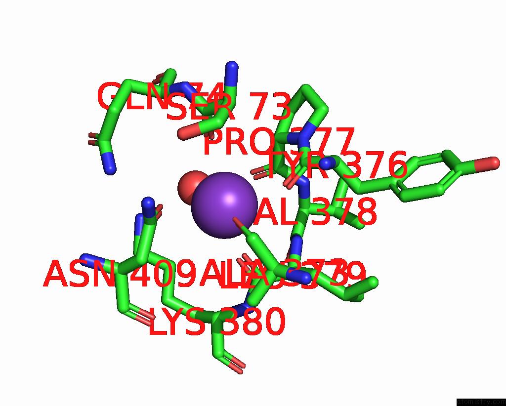
Mono view
Stereo pair view
Mono view
Stereo pair view
A full contact list of Potassium with other atoms in the K binding
site number 3 of Crystal Structure of Glucuronate Isomerase From Caulobacter Crescentus within 5.0Å range:
|
Reference:
Y.Patskovsky,
J.Bonanno,
V.Sridhar,
J.M.Sauder,
J.Freeman,
A.Powell,
J.Koss,
C.Groshong,
T.Gheyi,
S.R.Wasserman,
F.Raushel,
S.K.Burley,
S.C.Almo.
Crystal Structure of Glucuronate Isomerase From Caulobacter Crescentus. To Be Published.
Page generated: Sat Aug 9 03:47:27 2025
Last articles
Mn in 4UXAMn in 4W8Y
Mn in 4W9S
Mn in 4V15
Mn in 4V0U
Mn in 4V0W
Mn in 4V0X
Mn in 4UWQ
Mn in 4V0V
Mn in 4URR