Potassium »
PDB 2hzv-2o84 »
2i2x »
Potassium in PDB 2i2x: Crystal Structure of Methanol:Cobalamin Methyltransferase Complex Mtabc From Methanosarcina Barkeri
Enzymatic activity of Crystal Structure of Methanol:Cobalamin Methyltransferase Complex Mtabc From Methanosarcina Barkeri
All present enzymatic activity of Crystal Structure of Methanol:Cobalamin Methyltransferase Complex Mtabc From Methanosarcina Barkeri:
2.1.1.90;
2.1.1.90;
Protein crystallography data
The structure of Crystal Structure of Methanol:Cobalamin Methyltransferase Complex Mtabc From Methanosarcina Barkeri, PDB code: 2i2x
was solved by
C.H.Hagemeier,
M.Kruer,
R.K.Thauer,
E.Warkentin,
U.Ermler,
with X-Ray Crystallography technique. A brief refinement statistics is given in the table below:
| Resolution Low / High (Å) | 20.00 / 2.50 |
| Space group | P 1 21 1 |
| Cell size a, b, c (Å), α, β, γ (°) | 101.750, 172.850, 190.540, 90.00, 98.86, 90.00 |
| R / Rfree (%) | 18.2 / 23.1 |
Other elements in 2i2x:
The structure of Crystal Structure of Methanol:Cobalamin Methyltransferase Complex Mtabc From Methanosarcina Barkeri also contains other interesting chemical elements:
| Cobalt | (Co) | 8 atoms |
| Zinc | (Zn) | 12 atoms |
Potassium Binding Sites:
The binding sites of Potassium atom in the Crystal Structure of Methanol:Cobalamin Methyltransferase Complex Mtabc From Methanosarcina Barkeri
(pdb code 2i2x). This binding sites where shown within
5.0 Angstroms radius around Potassium atom.
In total 8 binding sites of Potassium where determined in the Crystal Structure of Methanol:Cobalamin Methyltransferase Complex Mtabc From Methanosarcina Barkeri, PDB code: 2i2x:
Jump to Potassium binding site number: 1; 2; 3; 4; 5; 6; 7; 8;
In total 8 binding sites of Potassium where determined in the Crystal Structure of Methanol:Cobalamin Methyltransferase Complex Mtabc From Methanosarcina Barkeri, PDB code: 2i2x:
Jump to Potassium binding site number: 1; 2; 3; 4; 5; 6; 7; 8;
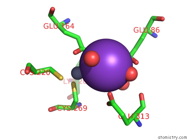
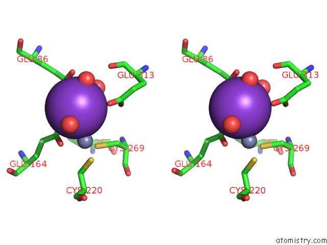
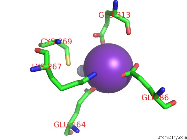
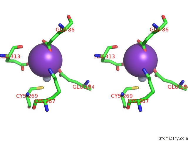
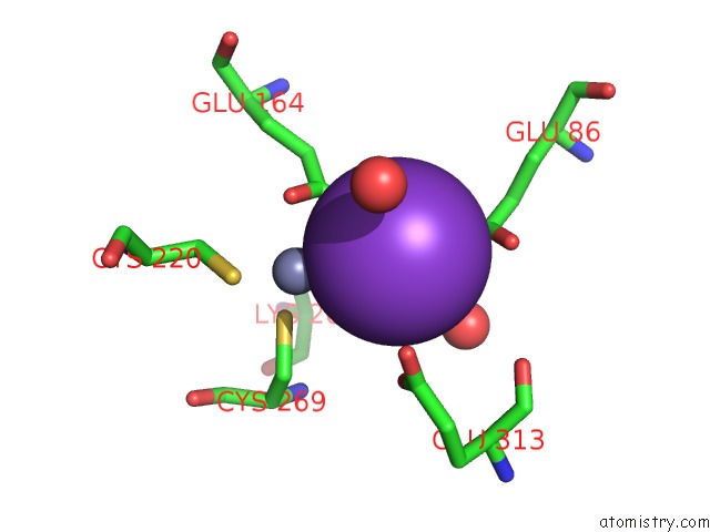
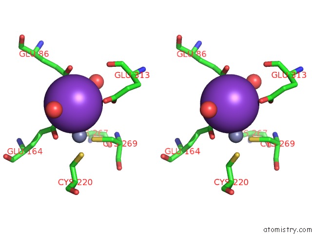
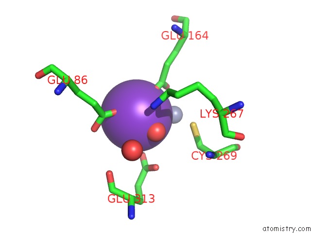
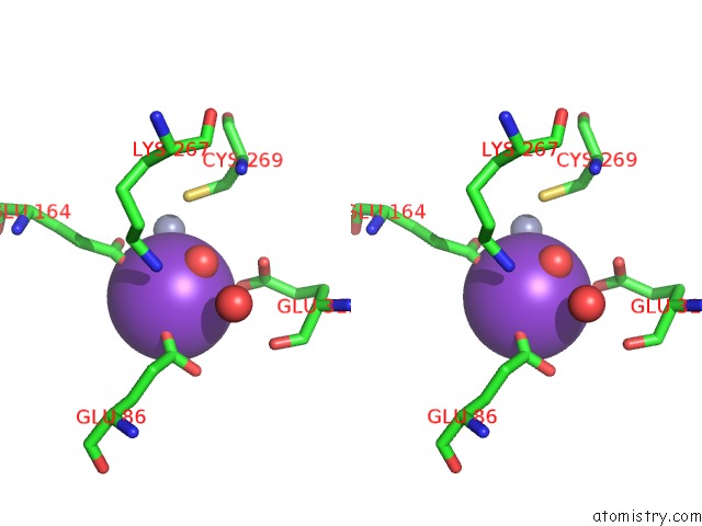
Potassium binding site 1 out of 8 in 2i2x
Go back to
Potassium binding site 1 out
of 8 in the Crystal Structure of Methanol:Cobalamin Methyltransferase Complex Mtabc From Methanosarcina Barkeri
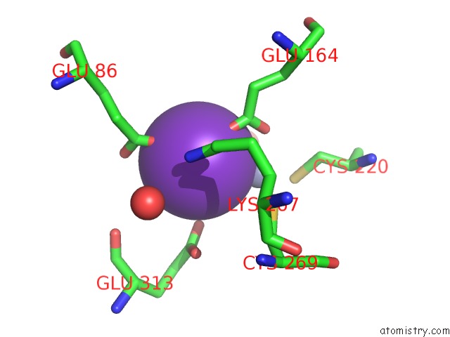
Mono view
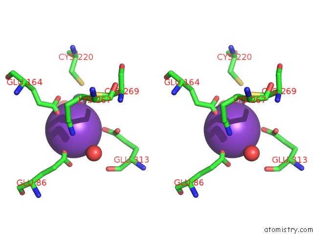
Stereo pair view
Mono view
Stereo pair view
A full contact list of Potassium with other atoms in the K binding
site number 1 of Crystal Structure of Methanol:Cobalamin Methyltransferase Complex Mtabc From Methanosarcina Barkeri within 5.0Å range:
|
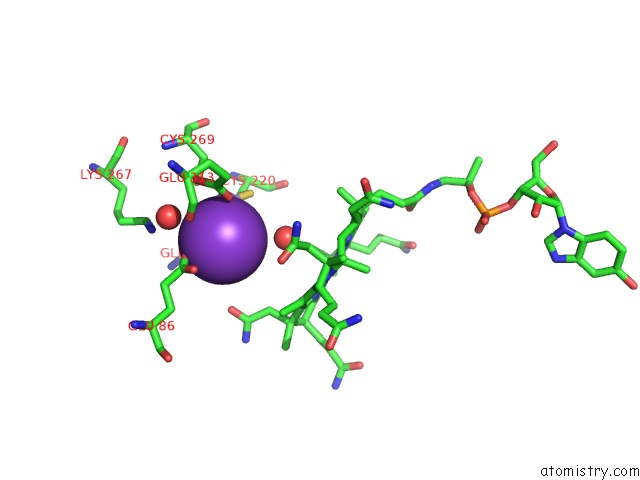
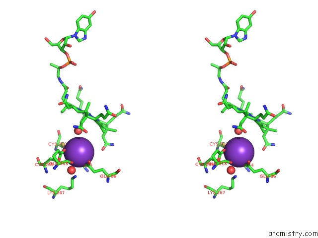
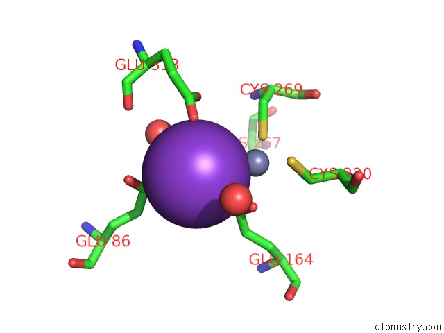
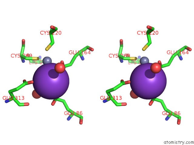
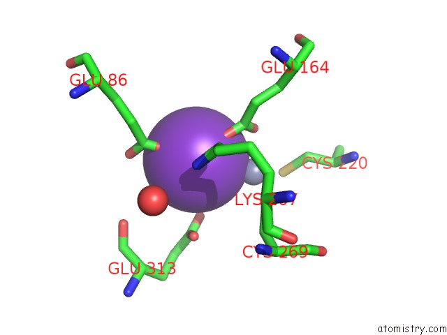
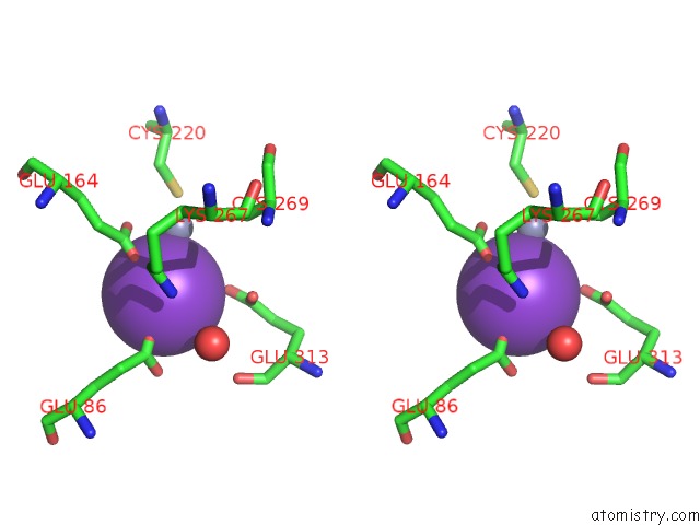
Potassium binding site 2 out of 8 in 2i2x
Go back to
Potassium binding site 2 out
of 8 in the Crystal Structure of Methanol:Cobalamin Methyltransferase Complex Mtabc From Methanosarcina Barkeri
Mono view
Stereo pair view
Mono view
Stereo pair view
A full contact list of Potassium with other atoms in the K binding
site number 2 of Crystal Structure of Methanol:Cobalamin Methyltransferase Complex Mtabc From Methanosarcina Barkeri within 5.0Å range:
|
Potassium binding site 3 out of 8 in 2i2x
Go back to
Potassium binding site 3 out
of 8 in the Crystal Structure of Methanol:Cobalamin Methyltransferase Complex Mtabc From Methanosarcina Barkeri
Mono view
Stereo pair view
Mono view
Stereo pair view
A full contact list of Potassium with other atoms in the K binding
site number 3 of Crystal Structure of Methanol:Cobalamin Methyltransferase Complex Mtabc From Methanosarcina Barkeri within 5.0Å range:
|
Potassium binding site 4 out of 8 in 2i2x
Go back to
Potassium binding site 4 out
of 8 in the Crystal Structure of Methanol:Cobalamin Methyltransferase Complex Mtabc From Methanosarcina Barkeri
Mono view
Stereo pair view
Mono view
Stereo pair view
A full contact list of Potassium with other atoms in the K binding
site number 4 of Crystal Structure of Methanol:Cobalamin Methyltransferase Complex Mtabc From Methanosarcina Barkeri within 5.0Å range:
|
Potassium binding site 5 out of 8 in 2i2x
Go back to
Potassium binding site 5 out
of 8 in the Crystal Structure of Methanol:Cobalamin Methyltransferase Complex Mtabc From Methanosarcina Barkeri
Mono view
Stereo pair view
Mono view
Stereo pair view
A full contact list of Potassium with other atoms in the K binding
site number 5 of Crystal Structure of Methanol:Cobalamin Methyltransferase Complex Mtabc From Methanosarcina Barkeri within 5.0Å range:
|
Potassium binding site 6 out of 8 in 2i2x
Go back to
Potassium binding site 6 out
of 8 in the Crystal Structure of Methanol:Cobalamin Methyltransferase Complex Mtabc From Methanosarcina Barkeri
Mono view
Stereo pair view
Mono view
Stereo pair view
A full contact list of Potassium with other atoms in the K binding
site number 6 of Crystal Structure of Methanol:Cobalamin Methyltransferase Complex Mtabc From Methanosarcina Barkeri within 5.0Å range:
|
Potassium binding site 7 out of 8 in 2i2x
Go back to
Potassium binding site 7 out
of 8 in the Crystal Structure of Methanol:Cobalamin Methyltransferase Complex Mtabc From Methanosarcina Barkeri
Mono view
Stereo pair view
Mono view
Stereo pair view
A full contact list of Potassium with other atoms in the K binding
site number 7 of Crystal Structure of Methanol:Cobalamin Methyltransferase Complex Mtabc From Methanosarcina Barkeri within 5.0Å range:
|
Potassium binding site 8 out of 8 in 2i2x
Go back to
Potassium binding site 8 out
of 8 in the Crystal Structure of Methanol:Cobalamin Methyltransferase Complex Mtabc From Methanosarcina Barkeri
Mono view
Stereo pair view
Mono view
Stereo pair view
A full contact list of Potassium with other atoms in the K binding
site number 8 of Crystal Structure of Methanol:Cobalamin Methyltransferase Complex Mtabc From Methanosarcina Barkeri within 5.0Å range:
|
Reference:
C.H.Hagemeier,
M.Krer,
R.K.Thauer,
E.Warkentin,
U.Ermler.
Insight Into the Mechanism of Biological Methanol Activation Based on the Crystal Structure of the Methanol-Cobalamin Methyltransferase Complex Proc.Natl.Acad.Sci.Usa V. 103 18917 2006.
ISSN: ISSN 0027-8424
PubMed: 17142327
DOI: 10.1073/PNAS.0603650103
Page generated: Sat Aug 9 03:36:52 2025
ISSN: ISSN 0027-8424
PubMed: 17142327
DOI: 10.1073/PNAS.0603650103
Last articles
Mg in 2A6EMg in 2A5Z
Mg in 2A5L
Mg in 2A5Y
Mg in 2A5J
Mg in 2A43
Mg in 2A5G
Mg in 2A5D
Mg in 2A5F
Mg in 2A42