Potassium »
PDB 2c44-2frz »
2dpb »
Potassium in PDB 2dpb: Crystal Structure of D(Cgcgaatxcgcg) Where X Is 5-(N- Aminohexyl)Carbamoyl-2'-Deoxyuridine
Protein crystallography data
The structure of Crystal Structure of D(Cgcgaatxcgcg) Where X Is 5-(N- Aminohexyl)Carbamoyl-2'-Deoxyuridine, PDB code: 2dpb
was solved by
E.C.M.Juan,
J.Kondo,
T.Kurihara,
T.Ito,
Y.Ueno,
A.Matsuda,
A.Takenaka,
with X-Ray Crystallography technique. A brief refinement statistics is given in the table below:
| Resolution Low / High (Å) | 10.00 / 1.50 |
| Space group | P 21 21 21 |
| Cell size a, b, c (Å), α, β, γ (°) | 25.539, 40.833, 64.456, 90.00, 90.00, 90.00 |
| R / Rfree (%) | 19.3 / 24.5 |
Potassium Binding Sites:
The binding sites of Potassium atom in the Crystal Structure of D(Cgcgaatxcgcg) Where X Is 5-(N- Aminohexyl)Carbamoyl-2'-Deoxyuridine
(pdb code 2dpb). This binding sites where shown within
5.0 Angstroms radius around Potassium atom.
In total only one binding site of Potassium was determined in the Crystal Structure of D(Cgcgaatxcgcg) Where X Is 5-(N- Aminohexyl)Carbamoyl-2'-Deoxyuridine, PDB code: 2dpb:
In total only one binding site of Potassium was determined in the Crystal Structure of D(Cgcgaatxcgcg) Where X Is 5-(N- Aminohexyl)Carbamoyl-2'-Deoxyuridine, PDB code: 2dpb:
Potassium binding site 1 out of 1 in 2dpb
Go back to
Potassium binding site 1 out
of 1 in the Crystal Structure of D(Cgcgaatxcgcg) Where X Is 5-(N- Aminohexyl)Carbamoyl-2'-Deoxyuridine
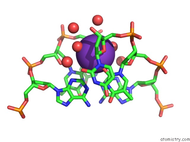
Mono view
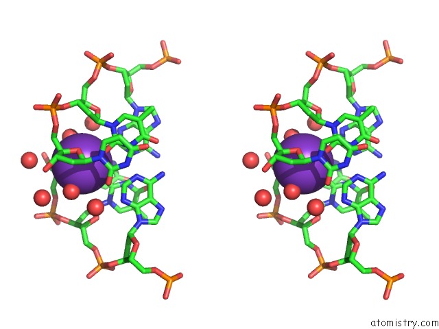
Stereo pair view
Mono view
Stereo pair view
A full contact list of Potassium with other atoms in the K binding
site number 1 of Crystal Structure of D(Cgcgaatxcgcg) Where X Is 5-(N- Aminohexyl)Carbamoyl-2'-Deoxyuridine within 5.0Å range:
|
Reference:
E.C.M.Juan,
J.Kondo,
T.Kurihara,
T.Ito,
Y.Ueno,
A.Matsuda,
A.Takenaka.
Crystal Structures of Dna:Dna and Dna:Rna Duplexes Containing 5-(N-Aminohexyl)Carbamoyl-Modified Uracils Reveal the Basis For Properties As Antigene and Antisense Molecules Nucleic Acids Res. V. 35 1969 2007.
ISSN: ISSN 0305-1048
PubMed: 17341465
DOI: 10.1093/NAR/GKL821
Page generated: Sat Aug 9 03:19:37 2025
ISSN: ISSN 0305-1048
PubMed: 17341465
DOI: 10.1093/NAR/GKL821
Last articles
Na in 3CCONa in 3CC2
Na in 3CCE
Na in 3CC7
Na in 3CC4
Na in 3CC9
Na in 3CBC
Na in 3CBT
Na in 3C9F
Na in 3CB8