Potassium »
PDB 2adp-2c13 »
2b5w »
Potassium in PDB 2b5w: Crystal Structure of D38C Glucose Dehydrogenase Mutant From Haloferax Mediterranei
Enzymatic activity of Crystal Structure of D38C Glucose Dehydrogenase Mutant From Haloferax Mediterranei
All present enzymatic activity of Crystal Structure of D38C Glucose Dehydrogenase Mutant From Haloferax Mediterranei:
1.1.1.47;
1.1.1.47;
Protein crystallography data
The structure of Crystal Structure of D38C Glucose Dehydrogenase Mutant From Haloferax Mediterranei, PDB code: 2b5w
was solved by
K.L.Britton,
P.J.Baker,
M.Fisher,
S.Ruzheinikov,
D.J.Gilmour,
M.-J.Bonete,
J.Ferrer,
C.Pire,
J.Esclapez,
D.W.Rice,
with X-Ray Crystallography technique. A brief refinement statistics is given in the table below:
| Resolution Low / High (Å) | 20.00 / 1.60 |
| Space group | I 2 2 2 |
| Cell size a, b, c (Å), α, β, γ (°) | 60.534, 109.255, 151.893, 90.00, 90.00, 90.00 |
| R / Rfree (%) | 15.4 / 18.6 |
Other elements in 2b5w:
The structure of Crystal Structure of D38C Glucose Dehydrogenase Mutant From Haloferax Mediterranei also contains other interesting chemical elements:
| Zinc | (Zn) | 1 atom |
Potassium Binding Sites:
The binding sites of Potassium atom in the Crystal Structure of D38C Glucose Dehydrogenase Mutant From Haloferax Mediterranei
(pdb code 2b5w). This binding sites where shown within
5.0 Angstroms radius around Potassium atom.
In total 5 binding sites of Potassium where determined in the Crystal Structure of D38C Glucose Dehydrogenase Mutant From Haloferax Mediterranei, PDB code: 2b5w:
Jump to Potassium binding site number: 1; 2; 3; 4; 5;
In total 5 binding sites of Potassium where determined in the Crystal Structure of D38C Glucose Dehydrogenase Mutant From Haloferax Mediterranei, PDB code: 2b5w:
Jump to Potassium binding site number: 1; 2; 3; 4; 5;
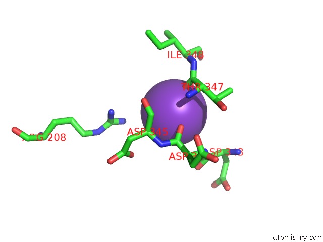
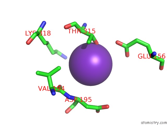
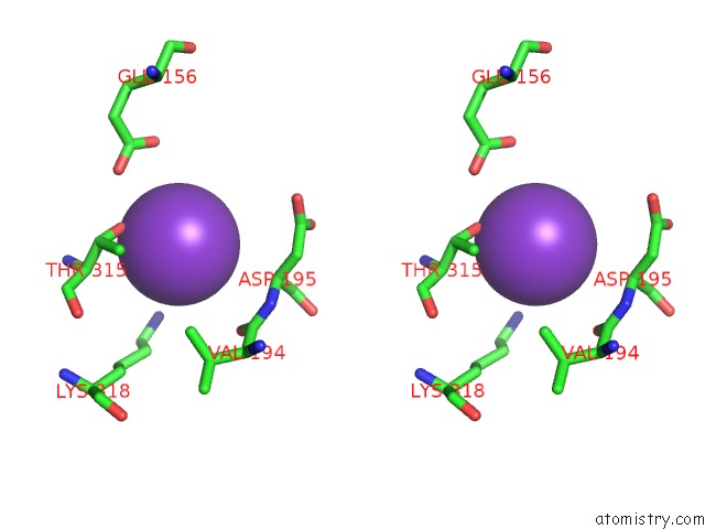
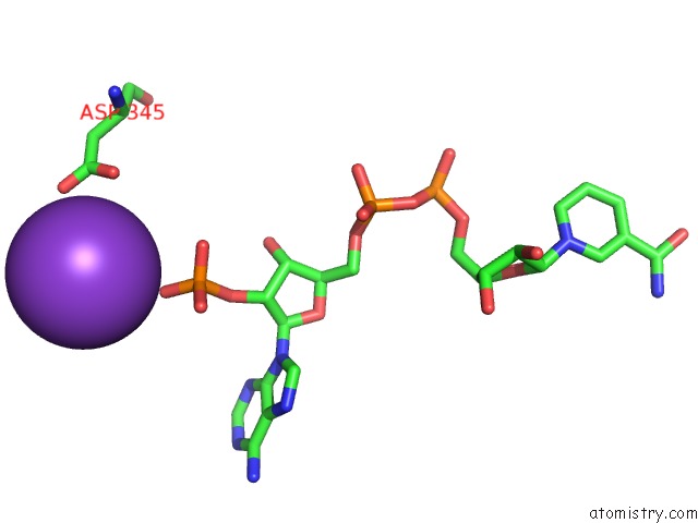
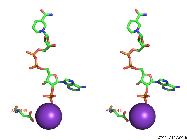
Potassium binding site 1 out of 5 in 2b5w
Go back to
Potassium binding site 1 out
of 5 in the Crystal Structure of D38C Glucose Dehydrogenase Mutant From Haloferax Mediterranei
Mono view
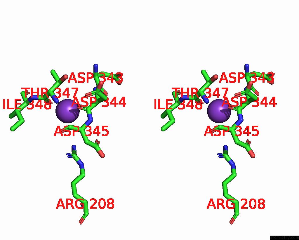
Stereo pair view
Mono view
Stereo pair view
A full contact list of Potassium with other atoms in the K binding
site number 1 of Crystal Structure of D38C Glucose Dehydrogenase Mutant From Haloferax Mediterranei within 5.0Å range:
|
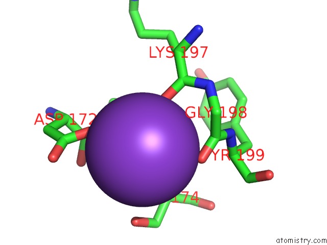
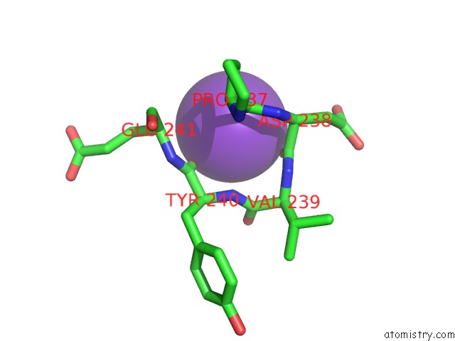
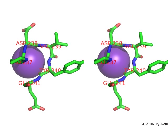
Potassium binding site 2 out of 5 in 2b5w
Go back to
Potassium binding site 2 out
of 5 in the Crystal Structure of D38C Glucose Dehydrogenase Mutant From Haloferax Mediterranei
Mono view
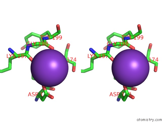
Stereo pair view
Mono view
Stereo pair view
A full contact list of Potassium with other atoms in the K binding
site number 2 of Crystal Structure of D38C Glucose Dehydrogenase Mutant From Haloferax Mediterranei within 5.0Å range:
|
Potassium binding site 3 out of 5 in 2b5w
Go back to
Potassium binding site 3 out
of 5 in the Crystal Structure of D38C Glucose Dehydrogenase Mutant From Haloferax Mediterranei
Mono view
Stereo pair view
Mono view
Stereo pair view
A full contact list of Potassium with other atoms in the K binding
site number 3 of Crystal Structure of D38C Glucose Dehydrogenase Mutant From Haloferax Mediterranei within 5.0Å range:
|
Potassium binding site 4 out of 5 in 2b5w
Go back to
Potassium binding site 4 out
of 5 in the Crystal Structure of D38C Glucose Dehydrogenase Mutant From Haloferax Mediterranei
Mono view
Stereo pair view
Mono view
Stereo pair view
A full contact list of Potassium with other atoms in the K binding
site number 4 of Crystal Structure of D38C Glucose Dehydrogenase Mutant From Haloferax Mediterranei within 5.0Å range:
|
Potassium binding site 5 out of 5 in 2b5w
Go back to
Potassium binding site 5 out
of 5 in the Crystal Structure of D38C Glucose Dehydrogenase Mutant From Haloferax Mediterranei
Mono view
Stereo pair view
Mono view
Stereo pair view
A full contact list of Potassium with other atoms in the K binding
site number 5 of Crystal Structure of D38C Glucose Dehydrogenase Mutant From Haloferax Mediterranei within 5.0Å range:
|
Reference:
K.L.Britton,
P.J.Baker,
M.Fisher,
S.Ruzheinikov,
D.J.Gilmour,
M.-J.Bonete,
J.Ferrer,
C.Pire,
J.Esclapez,
D.W.Rice.
Analysis of Protein Solvent Interactions in Glucose Dehydrogenase From the Extreme Halophile Haloferax Mediterranei. Proc.Natl.Acad.Sci.Usa V. 103 4846 2006.
ISSN: ISSN 0027-8424
PubMed: 16551747
DOI: 10.1073/PNAS.0508854103
Page generated: Sat Aug 9 03:11:54 2025
ISSN: ISSN 0027-8424
PubMed: 16551747
DOI: 10.1073/PNAS.0508854103
Last articles
Mg in 9DUFMg in 9DUD
Mg in 9DU8
Mg in 9DU9
Mg in 9DSZ
Mg in 9DUA
Mg in 9DTC
Mg in 9DTR
Mg in 9DTV
Mg in 9DT7