Potassium »
PDB 1yjn-2aaq »
2a6x »
Potassium in PDB 2a6x: Crystal Structure of EMP46P Carbohydrate Recognition Domain (Crd), Y131F Mutant
Protein crystallography data
The structure of Crystal Structure of EMP46P Carbohydrate Recognition Domain (Crd), Y131F Mutant, PDB code: 2a6x
was solved by
T.Satoh,
K.Sato,
A.Kanoh,
K.Yamashita,
R.Kato,
A.Nakano,
S.Wakatsuki,
Riken Structural Genomics/Proteomics Initiative(Rsgi),
with X-Ray Crystallography technique. A brief refinement statistics is given in the table below:
| Resolution Low / High (Å) | 20.00 / 1.55 |
| Space group | P 1 21 1 |
| Cell size a, b, c (Å), α, β, γ (°) | 54.240, 56.030, 77.670, 90.00, 108.64, 90.00 |
| R / Rfree (%) | 20.2 / 23.5 |
Potassium Binding Sites:
The binding sites of Potassium atom in the Crystal Structure of EMP46P Carbohydrate Recognition Domain (Crd), Y131F Mutant
(pdb code 2a6x). This binding sites where shown within
5.0 Angstroms radius around Potassium atom.
In total only one binding site of Potassium was determined in the Crystal Structure of EMP46P Carbohydrate Recognition Domain (Crd), Y131F Mutant, PDB code: 2a6x:
In total only one binding site of Potassium was determined in the Crystal Structure of EMP46P Carbohydrate Recognition Domain (Crd), Y131F Mutant, PDB code: 2a6x:
Potassium binding site 1 out of 1 in 2a6x
Go back to
Potassium binding site 1 out
of 1 in the Crystal Structure of EMP46P Carbohydrate Recognition Domain (Crd), Y131F Mutant

Mono view
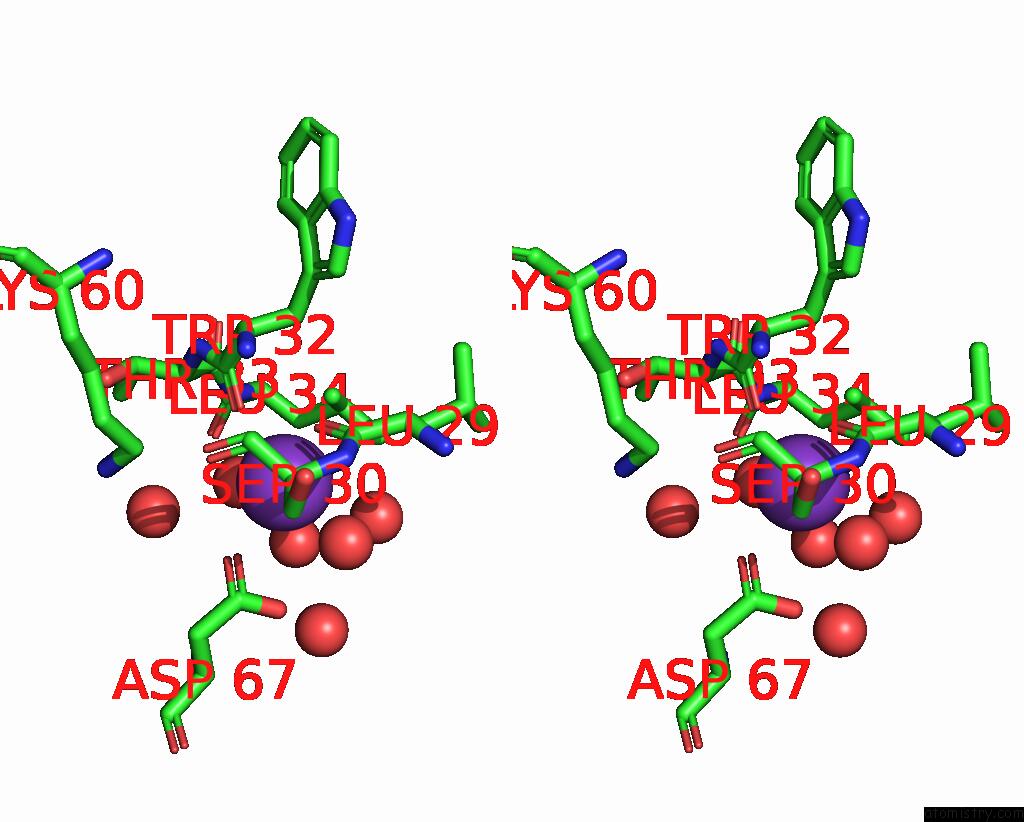
Stereo pair view
Mono view
Stereo pair view
A full contact list of Potassium with other atoms in the K binding
site number 1 of Crystal Structure of EMP46P Carbohydrate Recognition Domain (Crd), Y131F Mutant within 5.0Å range:
|
Reference:
T.Satoh,
K.Sato,
A.Kanoh,
K.Yamashita,
Y.Yamada,
N.Igarashi,
R.Kato,
A.Nakano,
S.Wakatsuki.
Structures of the Carbohydrate Recognition Domain of CA2+-Independent Cargo Receptors EMP46P and EMP47P. J.Biol.Chem. V. 281 10410 2006.
ISSN: ISSN 0021-9258
PubMed: 16439369
DOI: 10.1074/JBC.M512258200
Page generated: Sat Aug 9 03:06:07 2025
ISSN: ISSN 0021-9258
PubMed: 16439369
DOI: 10.1074/JBC.M512258200
Last articles
Na in 8H8TNa in 8H8Q
Na in 8H58
Na in 8H8P
Na in 8H7N
Na in 8H63
Na in 8GT0
Na in 8H2B
Na in 8GZ1
Na in 8H1G