Potassium »
PDB 1yjn-2aaq »
2a1m »
Potassium in PDB 2a1m: Crystal Structure of Ferrous Dioxygen Complex of Wild-Type Cytochrome P450CAM
Enzymatic activity of Crystal Structure of Ferrous Dioxygen Complex of Wild-Type Cytochrome P450CAM
All present enzymatic activity of Crystal Structure of Ferrous Dioxygen Complex of Wild-Type Cytochrome P450CAM:
1.14.15.1;
1.14.15.1;
Protein crystallography data
The structure of Crystal Structure of Ferrous Dioxygen Complex of Wild-Type Cytochrome P450CAM, PDB code: 2a1m
was solved by
S.Nagano,
T.L.Poulos,
with X-Ray Crystallography technique. A brief refinement statistics is given in the table below:
| Resolution Low / High (Å) | 47.82 / 2.10 |
| Space group | P 1 21 1 |
| Cell size a, b, c (Å), α, β, γ (°) | 67.060, 62.290, 95.640, 90.00, 90.42, 90.00 |
| R / Rfree (%) | 19.1 / 24.7 |
Other elements in 2a1m:
The structure of Crystal Structure of Ferrous Dioxygen Complex of Wild-Type Cytochrome P450CAM also contains other interesting chemical elements:
| Iron | (Fe) | 2 atoms |
Potassium Binding Sites:
The binding sites of Potassium atom in the Crystal Structure of Ferrous Dioxygen Complex of Wild-Type Cytochrome P450CAM
(pdb code 2a1m). This binding sites where shown within
5.0 Angstroms radius around Potassium atom.
In total 3 binding sites of Potassium where determined in the Crystal Structure of Ferrous Dioxygen Complex of Wild-Type Cytochrome P450CAM, PDB code: 2a1m:
Jump to Potassium binding site number: 1; 2; 3;
In total 3 binding sites of Potassium where determined in the Crystal Structure of Ferrous Dioxygen Complex of Wild-Type Cytochrome P450CAM, PDB code: 2a1m:
Jump to Potassium binding site number: 1; 2; 3;
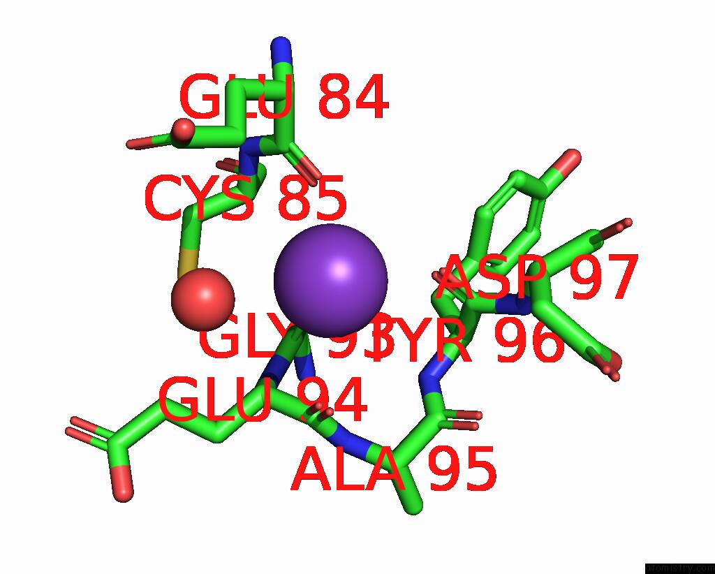
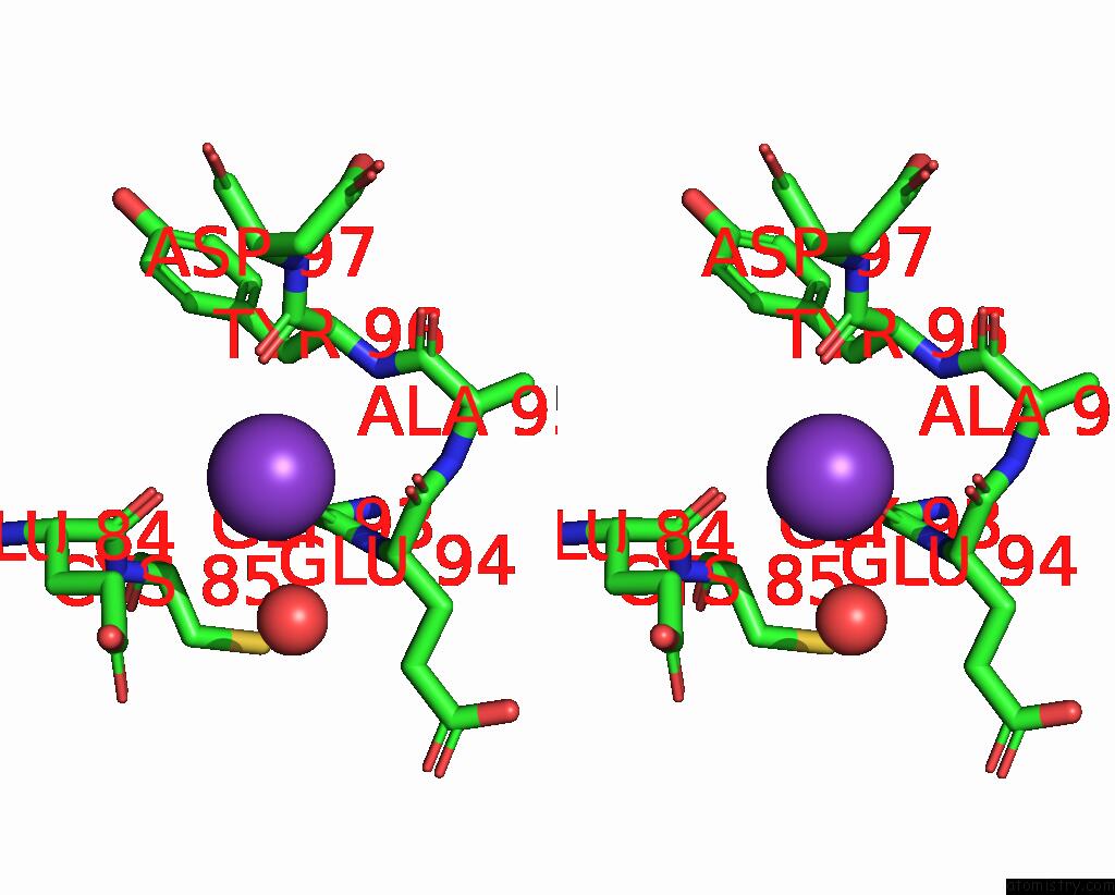
Potassium binding site 1 out of 3 in 2a1m
Go back to
Potassium binding site 1 out
of 3 in the Crystal Structure of Ferrous Dioxygen Complex of Wild-Type Cytochrome P450CAM
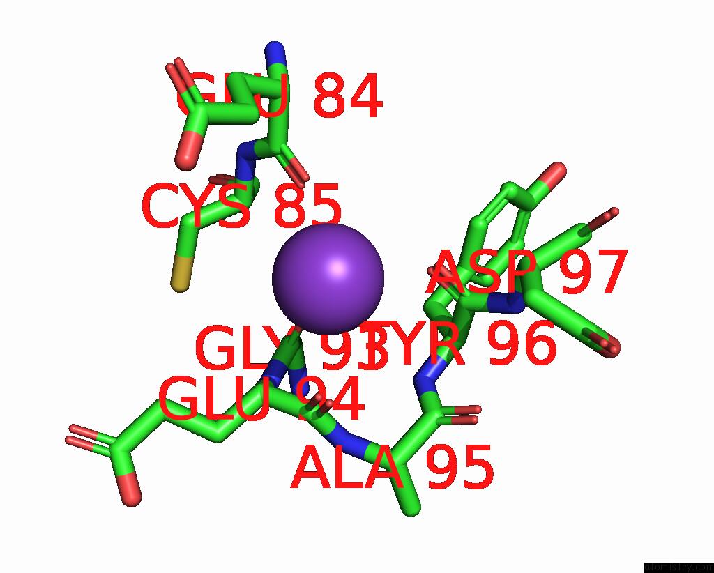
Mono view

Stereo pair view
Mono view
Stereo pair view
A full contact list of Potassium with other atoms in the K binding
site number 1 of Crystal Structure of Ferrous Dioxygen Complex of Wild-Type Cytochrome P450CAM within 5.0Å range:
|
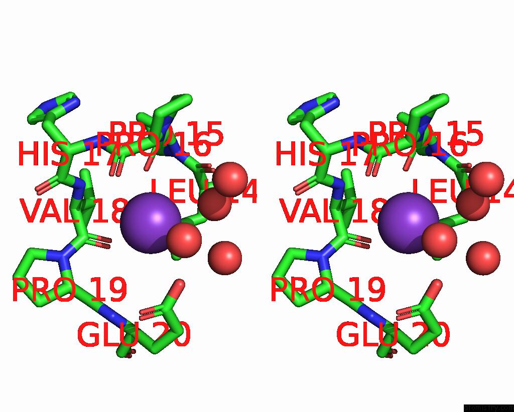
Potassium binding site 2 out of 3 in 2a1m
Go back to
Potassium binding site 2 out
of 3 in the Crystal Structure of Ferrous Dioxygen Complex of Wild-Type Cytochrome P450CAM
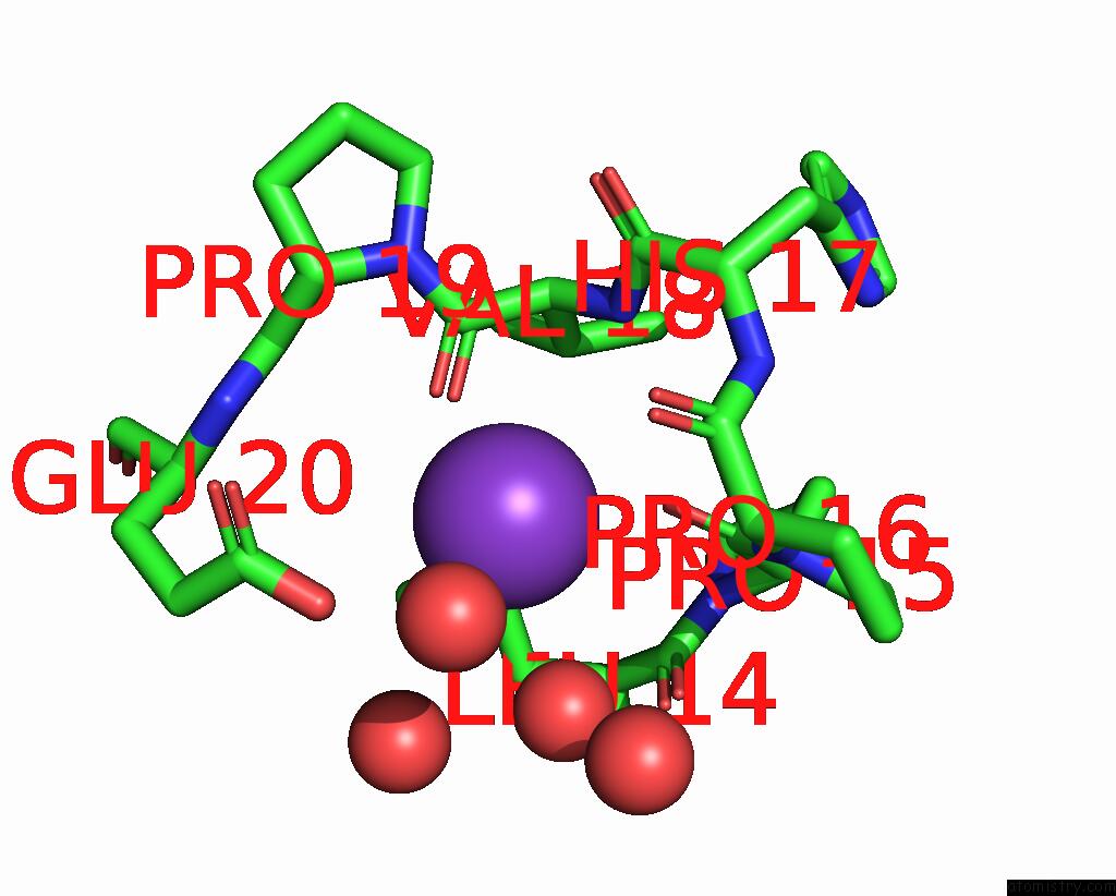
Mono view
Stereo pair view
Mono view
Stereo pair view
A full contact list of Potassium with other atoms in the K binding
site number 2 of Crystal Structure of Ferrous Dioxygen Complex of Wild-Type Cytochrome P450CAM within 5.0Å range:
|
Potassium binding site 3 out of 3 in 2a1m
Go back to
Potassium binding site 3 out
of 3 in the Crystal Structure of Ferrous Dioxygen Complex of Wild-Type Cytochrome P450CAM
Mono view
Stereo pair view
Mono view
Stereo pair view
A full contact list of Potassium with other atoms in the K binding
site number 3 of Crystal Structure of Ferrous Dioxygen Complex of Wild-Type Cytochrome P450CAM within 5.0Å range:
|
Reference:
S.Nagano,
T.L.Poulos.
Crystallographic Study on the Dioxygen Complex of Wild-Type and Mutant Cytochrome P450CAM. Implications For the Dioxygen Activation Mechanism J.Biol.Chem. V. 280 31659 2005.
ISSN: ISSN 0021-9258
PubMed: 15994329
DOI: 10.1074/JBC.M505261200
Page generated: Mon Aug 12 05:57:21 2024
ISSN: ISSN 0021-9258
PubMed: 15994329
DOI: 10.1074/JBC.M505261200
Last articles
F in 5JFRF in 5JEI
F in 5JAJ
F in 5JBG
F in 5JB2
F in 5J8U
F in 5J9X
F in 5JAL
F in 5J7P
F in 5J8M