Potassium »
PDB 1yjn-2aaq »
1zz0 »
Potassium in PDB 1zz0: Crystal Structure of A Hdac-Like Protein with Acetate Bound
Protein crystallography data
The structure of Crystal Structure of A Hdac-Like Protein with Acetate Bound, PDB code: 1zz0
was solved by
T.K.Nielsen,
C.Hildmann,
A.Dickmanns,
A.Schwienhorst,
R.Ficner,
with X-Ray Crystallography technique. A brief refinement statistics is given in the table below:
| Resolution Low / High (Å) | 44.86 / 1.60 |
| Space group | I 2 2 2 |
| Cell size a, b, c (Å), α, β, γ (°) | 93.586, 127.886, 251.681, 90.00, 90.00, 90.00 |
| R / Rfree (%) | 16.2 / 18.6 |
Other elements in 1zz0:
The structure of Crystal Structure of A Hdac-Like Protein with Acetate Bound also contains other interesting chemical elements:
| Zinc | (Zn) | 4 atoms |
Potassium Binding Sites:
The binding sites of Potassium atom in the Crystal Structure of A Hdac-Like Protein with Acetate Bound
(pdb code 1zz0). This binding sites where shown within
5.0 Angstroms radius around Potassium atom.
In total 8 binding sites of Potassium where determined in the Crystal Structure of A Hdac-Like Protein with Acetate Bound, PDB code: 1zz0:
Jump to Potassium binding site number: 1; 2; 3; 4; 5; 6; 7; 8;
In total 8 binding sites of Potassium where determined in the Crystal Structure of A Hdac-Like Protein with Acetate Bound, PDB code: 1zz0:
Jump to Potassium binding site number: 1; 2; 3; 4; 5; 6; 7; 8;
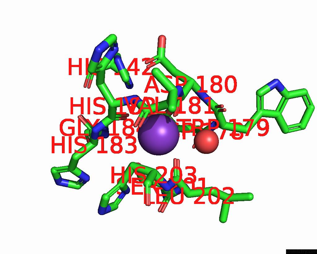
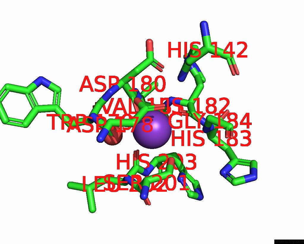
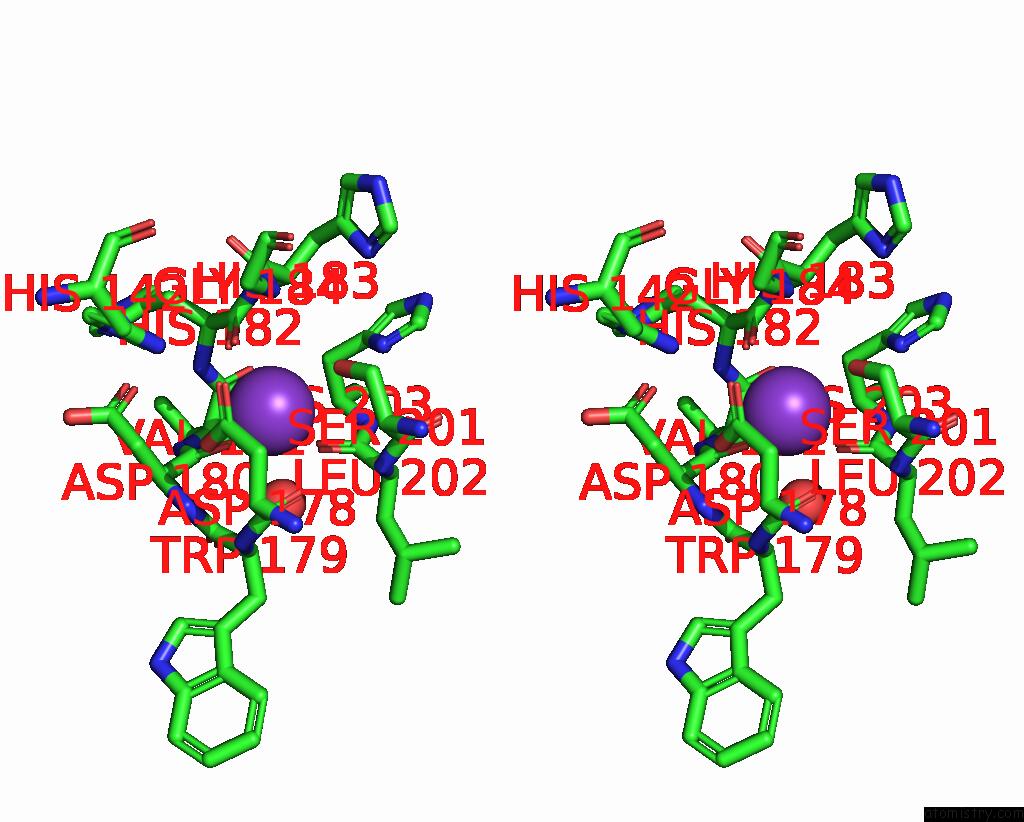
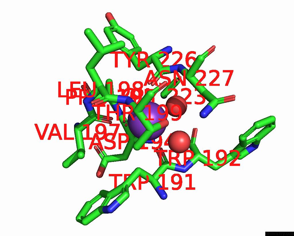
Potassium binding site 1 out of 8 in 1zz0
Go back to
Potassium binding site 1 out
of 8 in the Crystal Structure of A Hdac-Like Protein with Acetate Bound

Mono view
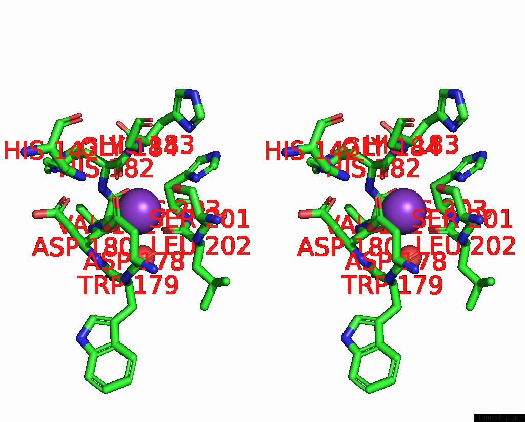
Stereo pair view
Mono view
Stereo pair view
A full contact list of Potassium with other atoms in the K binding
site number 1 of Crystal Structure of A Hdac-Like Protein with Acetate Bound within 5.0Å range:
|
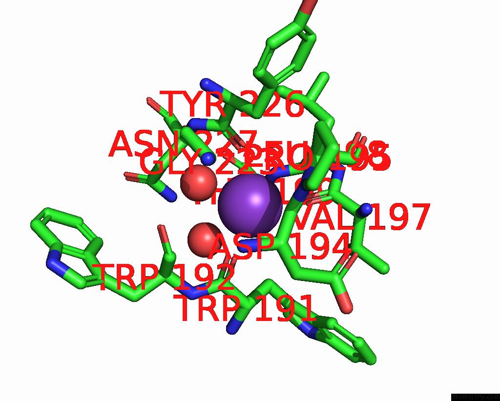
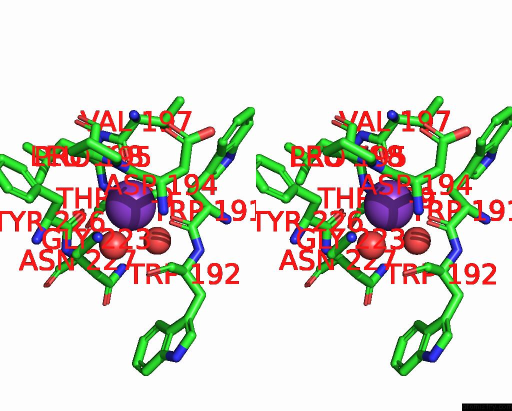
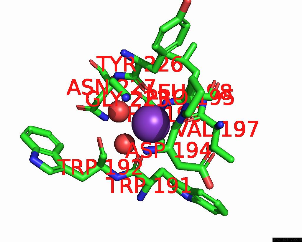
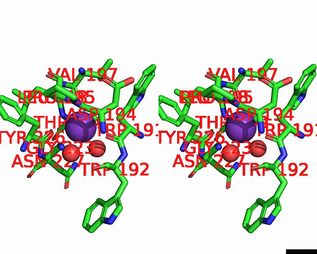
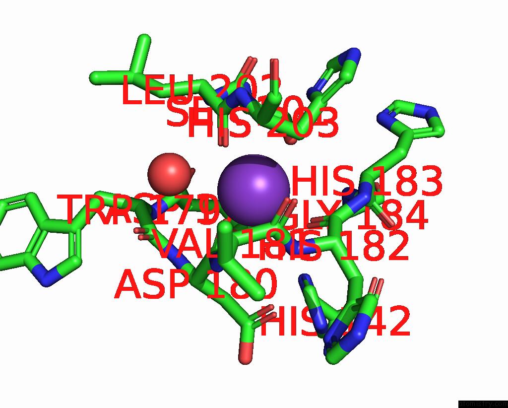
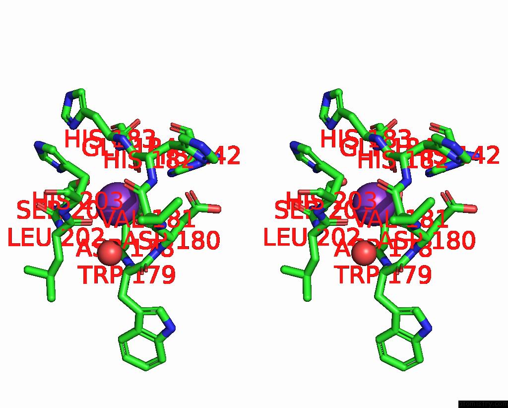
Potassium binding site 2 out of 8 in 1zz0
Go back to
Potassium binding site 2 out
of 8 in the Crystal Structure of A Hdac-Like Protein with Acetate Bound
Mono view
Stereo pair view
Mono view
Stereo pair view
A full contact list of Potassium with other atoms in the K binding
site number 2 of Crystal Structure of A Hdac-Like Protein with Acetate Bound within 5.0Å range:
|
Potassium binding site 3 out of 8 in 1zz0
Go back to
Potassium binding site 3 out
of 8 in the Crystal Structure of A Hdac-Like Protein with Acetate Bound
Mono view

Stereo pair view
Mono view
Stereo pair view
A full contact list of Potassium with other atoms in the K binding
site number 3 of Crystal Structure of A Hdac-Like Protein with Acetate Bound within 5.0Å range:
|
Potassium binding site 4 out of 8 in 1zz0
Go back to
Potassium binding site 4 out
of 8 in the Crystal Structure of A Hdac-Like Protein with Acetate Bound

Mono view

Stereo pair view
Mono view
Stereo pair view
A full contact list of Potassium with other atoms in the K binding
site number 4 of Crystal Structure of A Hdac-Like Protein with Acetate Bound within 5.0Å range:
|
Potassium binding site 5 out of 8 in 1zz0
Go back to
Potassium binding site 5 out
of 8 in the Crystal Structure of A Hdac-Like Protein with Acetate Bound
Mono view
Stereo pair view
Mono view
Stereo pair view
A full contact list of Potassium with other atoms in the K binding
site number 5 of Crystal Structure of A Hdac-Like Protein with Acetate Bound within 5.0Å range:
|
Potassium binding site 6 out of 8 in 1zz0
Go back to
Potassium binding site 6 out
of 8 in the Crystal Structure of A Hdac-Like Protein with Acetate Bound
Mono view
Stereo pair view
Mono view
Stereo pair view
A full contact list of Potassium with other atoms in the K binding
site number 6 of Crystal Structure of A Hdac-Like Protein with Acetate Bound within 5.0Å range:
|
Potassium binding site 7 out of 8 in 1zz0
Go back to
Potassium binding site 7 out
of 8 in the Crystal Structure of A Hdac-Like Protein with Acetate Bound
Mono view

Stereo pair view
Mono view
Stereo pair view
A full contact list of Potassium with other atoms in the K binding
site number 7 of Crystal Structure of A Hdac-Like Protein with Acetate Bound within 5.0Å range:
|
Potassium binding site 8 out of 8 in 1zz0
Go back to
Potassium binding site 8 out
of 8 in the Crystal Structure of A Hdac-Like Protein with Acetate Bound
Mono view
Stereo pair view
Mono view
Stereo pair view
A full contact list of Potassium with other atoms in the K binding
site number 8 of Crystal Structure of A Hdac-Like Protein with Acetate Bound within 5.0Å range:
|
Reference:
T.K.Nielsen,
C.Hildmann,
A.Dickmanns,
A.Schwienhorst,
R.Ficner.
Crystal Structure of A Bacterial Class 2 Histone Deacetylase Homologue J.Mol.Biol. V. 354 107 2005.
ISSN: ISSN 0022-2836
PubMed: 16242151
DOI: 10.1016/J.JMB.2005.09.065
Page generated: Sat Aug 9 03:02:16 2025
ISSN: ISSN 0022-2836
PubMed: 16242151
DOI: 10.1016/J.JMB.2005.09.065
Last articles
Na in 8GF8Na in 8GCQ
Na in 8GBT
Na in 8G8A
Na in 8G1Y
Na in 8G8V
Na in 8G62
Na in 8G2H
Na in 8G2E
Na in 8G1L