Potassium »
PDB 1yjn-2aaq »
1yrd »
Potassium in PDB 1yrd: X-Ray Crystal Structure of Perdeuterated Cytochrome P450CAM
Enzymatic activity of X-Ray Crystal Structure of Perdeuterated Cytochrome P450CAM
All present enzymatic activity of X-Ray Crystal Structure of Perdeuterated Cytochrome P450CAM:
1.14.15.1;
1.14.15.1;
Protein crystallography data
The structure of X-Ray Crystal Structure of Perdeuterated Cytochrome P450CAM, PDB code: 1yrd
was solved by
F.Meilleur,
M.-T.Dauvergne,
I.Schlichting,
D.A.A.Myles,
with X-Ray Crystallography technique. A brief refinement statistics is given in the table below:
| Resolution Low / High (Å) | 20.00 / 1.70 |
| Space group | P 43 21 2 |
| Cell size a, b, c (Å), α, β, γ (°) | 63.430, 63.430, 248.640, 90.00, 90.00, 90.00 |
| R / Rfree (%) | 19.8 / 21.3 |
Other elements in 1yrd:
The structure of X-Ray Crystal Structure of Perdeuterated Cytochrome P450CAM also contains other interesting chemical elements:
| Iron | (Fe) | 1 atom |
Potassium Binding Sites:
The binding sites of Potassium atom in the X-Ray Crystal Structure of Perdeuterated Cytochrome P450CAM
(pdb code 1yrd). This binding sites where shown within
5.0 Angstroms radius around Potassium atom.
In total only one binding site of Potassium was determined in the X-Ray Crystal Structure of Perdeuterated Cytochrome P450CAM, PDB code: 1yrd:
In total only one binding site of Potassium was determined in the X-Ray Crystal Structure of Perdeuterated Cytochrome P450CAM, PDB code: 1yrd:
Potassium binding site 1 out of 1 in 1yrd
Go back to
Potassium binding site 1 out
of 1 in the X-Ray Crystal Structure of Perdeuterated Cytochrome P450CAM
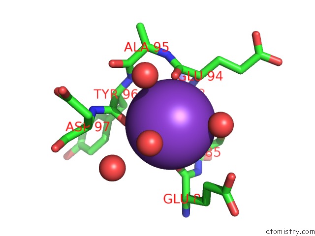
Mono view
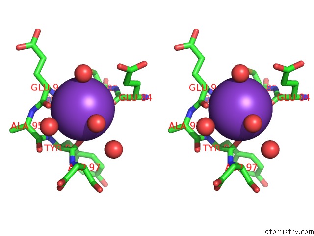
Stereo pair view
Mono view
Stereo pair view
A full contact list of Potassium with other atoms in the K binding
site number 1 of X-Ray Crystal Structure of Perdeuterated Cytochrome P450CAM within 5.0Å range:
|
Reference:
F.Meilleur,
M.T.Dauvergne,
I.Schlichting,
D.A.Myles.
Production and X-Ray Crystallographic Analysis of Fully Deuterated Cytochrome P450CAM. Acta Crystallogr.,Sect.D V. 61 539 2005.
ISSN: ISSN 0907-4449
PubMed: 15858263
DOI: 10.1107/S0907444905003872
Page generated: Sat Aug 9 02:59:52 2025
ISSN: ISSN 0907-4449
PubMed: 15858263
DOI: 10.1107/S0907444905003872
Last articles
Na in 8GMWNa in 8GN9
Na in 8GMV
Na in 8GKO
Na in 8GKM
Na in 8GLH
Na in 8GHF
Na in 8GI9
Na in 8GKA
Na in 8GI8