Potassium »
PDB 1u1g-1w29 »
1vko »
Potassium in PDB 1vko: Crystal Structure of Inositol-3-Phosphate Synthase (CE21227) From Caenorhabditis Elegans at 2.30 A Resolution
Enzymatic activity of Crystal Structure of Inositol-3-Phosphate Synthase (CE21227) From Caenorhabditis Elegans at 2.30 A Resolution
All present enzymatic activity of Crystal Structure of Inositol-3-Phosphate Synthase (CE21227) From Caenorhabditis Elegans at 2.30 A Resolution:
5.5.1.4;
5.5.1.4;
Protein crystallography data
The structure of Crystal Structure of Inositol-3-Phosphate Synthase (CE21227) From Caenorhabditis Elegans at 2.30 A Resolution, PDB code: 1vko
was solved by
Joint Center For Structural Genomics (Jcsg),
with X-Ray Crystallography technique. A brief refinement statistics is given in the table below:
| Resolution Low / High (Å) | 29.41 / 2.30 |
| Space group | I 2 2 2 |
| Cell size a, b, c (Å), α, β, γ (°) | 76.980, 129.980, 131.320, 90.00, 90.00, 90.00 |
| R / Rfree (%) | 20 / 26.5 |
Other elements in 1vko:
The structure of Crystal Structure of Inositol-3-Phosphate Synthase (CE21227) From Caenorhabditis Elegans at 2.30 A Resolution also contains other interesting chemical elements:
| Iodine | (I) | 2 atoms |
| Chlorine | (Cl) | 2 atoms |
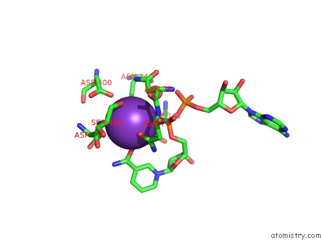
Potassium Binding Sites:
The binding sites of Potassium atom in the Crystal Structure of Inositol-3-Phosphate Synthase (CE21227) From Caenorhabditis Elegans at 2.30 A Resolution
(pdb code 1vko). This binding sites where shown within
5.0 Angstroms radius around Potassium atom.
In total only one binding site of Potassium was determined in the Crystal Structure of Inositol-3-Phosphate Synthase (CE21227) From Caenorhabditis Elegans at 2.30 A Resolution, PDB code: 1vko:
In total only one binding site of Potassium was determined in the Crystal Structure of Inositol-3-Phosphate Synthase (CE21227) From Caenorhabditis Elegans at 2.30 A Resolution, PDB code: 1vko:
Potassium binding site 1 out of 1 in 1vko
Go back to
Potassium binding site 1 out
of 1 in the Crystal Structure of Inositol-3-Phosphate Synthase (CE21227) From Caenorhabditis Elegans at 2.30 A Resolution
Mono view
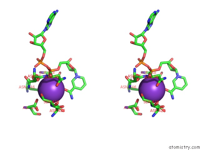
Stereo pair view
Mono view
Stereo pair view
A full contact list of Potassium with other atoms in the K binding
site number 1 of Crystal Structure of Inositol-3-Phosphate Synthase (CE21227) From Caenorhabditis Elegans at 2.30 A Resolution within 5.0Å range:
|
Reference:
Joint Center For Structural Genomics (Jcsg),
Joint Center For Structural Genomics (Jcsg).
N/A N/A.
Page generated: Sat Aug 9 02:47:37 2025
Last articles
Na in 8VMYNa in 8VMX
Na in 8VMW
Na in 8VL8
Na in 8VMU
Na in 8VMT
Na in 8VMV
Na in 8VMS
Na in 8VMR
Na in 8VMQ