Potassium »
PDB 1pr9-1s61 »
1pwh »
Potassium in PDB 1pwh: Rat Liver L-Serine Dehydratase- Complex with Pyridoxyl-(O-Methyl- Serine)-5-Monophosphate
Enzymatic activity of Rat Liver L-Serine Dehydratase- Complex with Pyridoxyl-(O-Methyl- Serine)-5-Monophosphate
All present enzymatic activity of Rat Liver L-Serine Dehydratase- Complex with Pyridoxyl-(O-Methyl- Serine)-5-Monophosphate:
4.3.1.17;
4.3.1.17;
Protein crystallography data
The structure of Rat Liver L-Serine Dehydratase- Complex with Pyridoxyl-(O-Methyl- Serine)-5-Monophosphate, PDB code: 1pwh
was solved by
T.Yamada,
J.Komoto,
Y.Takata,
H.Ogawa,
F.Takusagawa,
with X-Ray Crystallography technique. A brief refinement statistics is given in the table below:
| Resolution Low / High (Å) | 25.01 / 2.60 |
| Space group | P 1 21 1 |
| Cell size a, b, c (Å), α, β, γ (°) | 62.241, 109.262, 98.940, 90.00, 91.66, 90.00 |
| R / Rfree (%) | 23 / 26 |
Potassium Binding Sites:
The binding sites of Potassium atom in the Rat Liver L-Serine Dehydratase- Complex with Pyridoxyl-(O-Methyl- Serine)-5-Monophosphate
(pdb code 1pwh). This binding sites where shown within
5.0 Angstroms radius around Potassium atom.
In total 4 binding sites of Potassium where determined in the Rat Liver L-Serine Dehydratase- Complex with Pyridoxyl-(O-Methyl- Serine)-5-Monophosphate, PDB code: 1pwh:
Jump to Potassium binding site number: 1; 2; 3; 4;
In total 4 binding sites of Potassium where determined in the Rat Liver L-Serine Dehydratase- Complex with Pyridoxyl-(O-Methyl- Serine)-5-Monophosphate, PDB code: 1pwh:
Jump to Potassium binding site number: 1; 2; 3; 4;
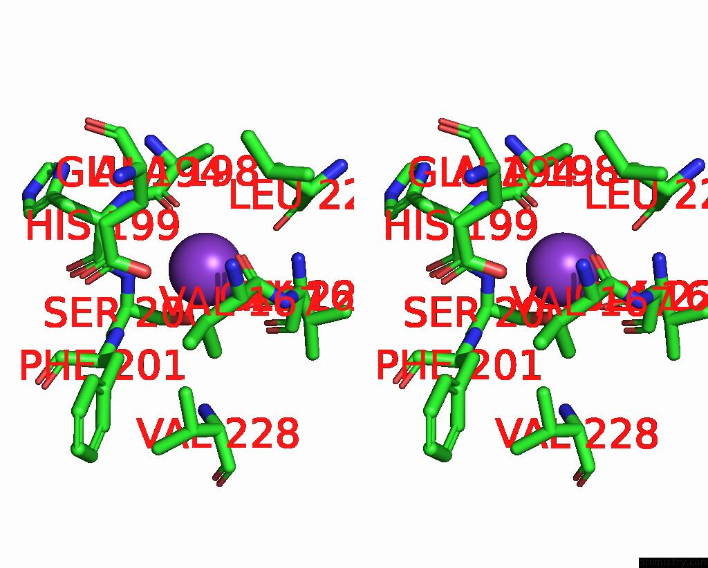
Potassium binding site 1 out of 4 in 1pwh
Go back to
Potassium binding site 1 out
of 4 in the Rat Liver L-Serine Dehydratase- Complex with Pyridoxyl-(O-Methyl- Serine)-5-Monophosphate
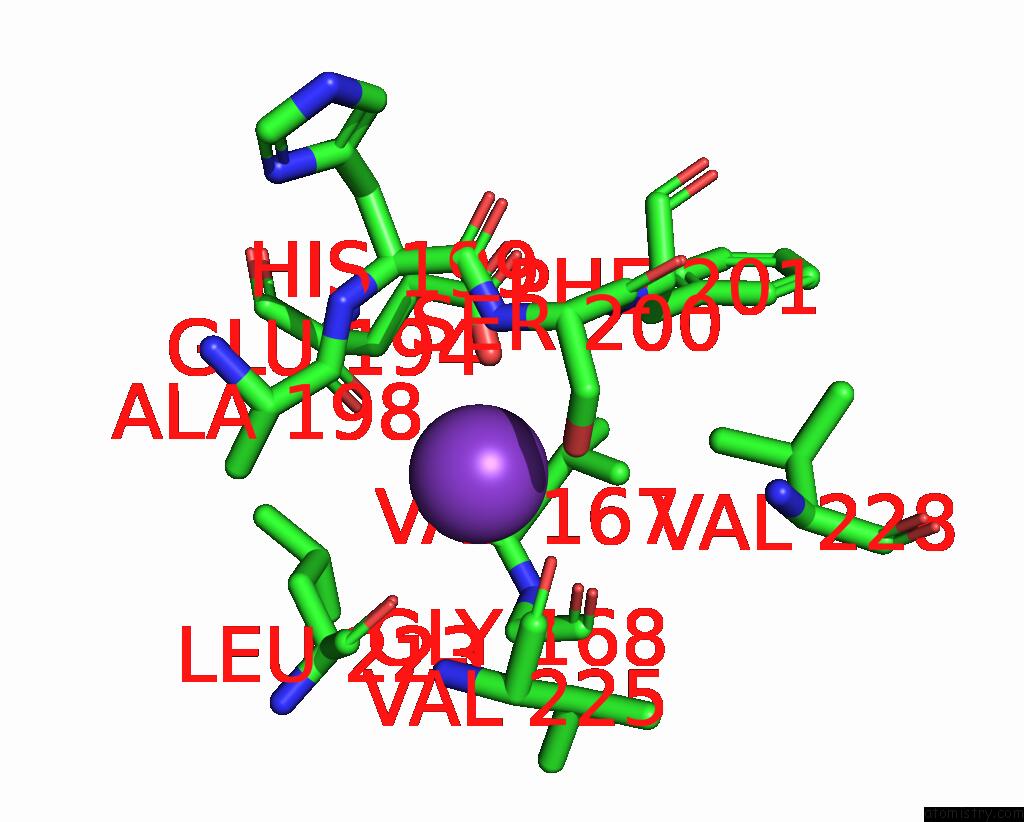
Mono view
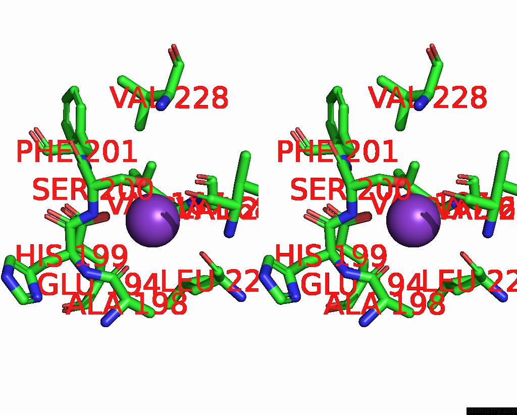
Stereo pair view
Mono view
Stereo pair view
A full contact list of Potassium with other atoms in the K binding
site number 1 of Rat Liver L-Serine Dehydratase- Complex with Pyridoxyl-(O-Methyl- Serine)-5-Monophosphate within 5.0Å range:
|
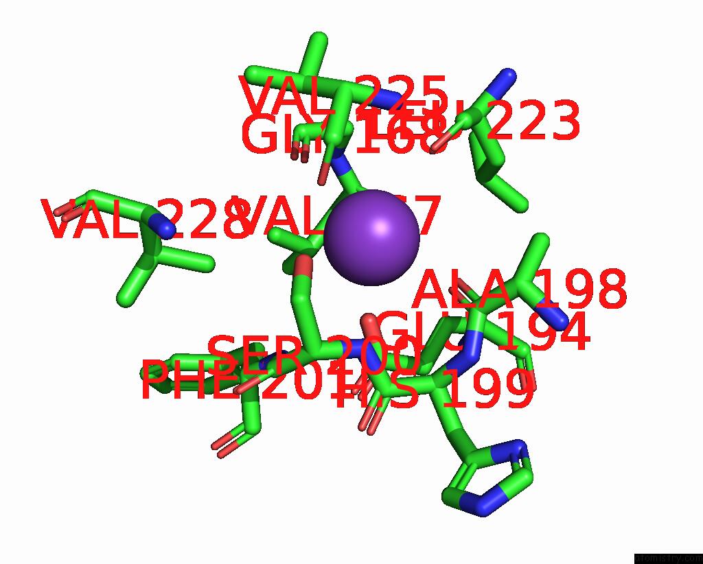
Potassium binding site 2 out of 4 in 1pwh
Go back to
Potassium binding site 2 out
of 4 in the Rat Liver L-Serine Dehydratase- Complex with Pyridoxyl-(O-Methyl- Serine)-5-Monophosphate
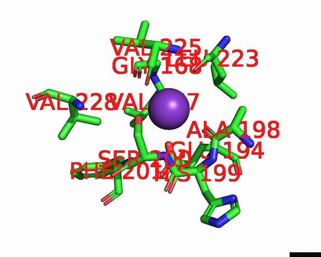
Mono view
Stereo pair view
Mono view
Stereo pair view
A full contact list of Potassium with other atoms in the K binding
site number 2 of Rat Liver L-Serine Dehydratase- Complex with Pyridoxyl-(O-Methyl- Serine)-5-Monophosphate within 5.0Å range:
|
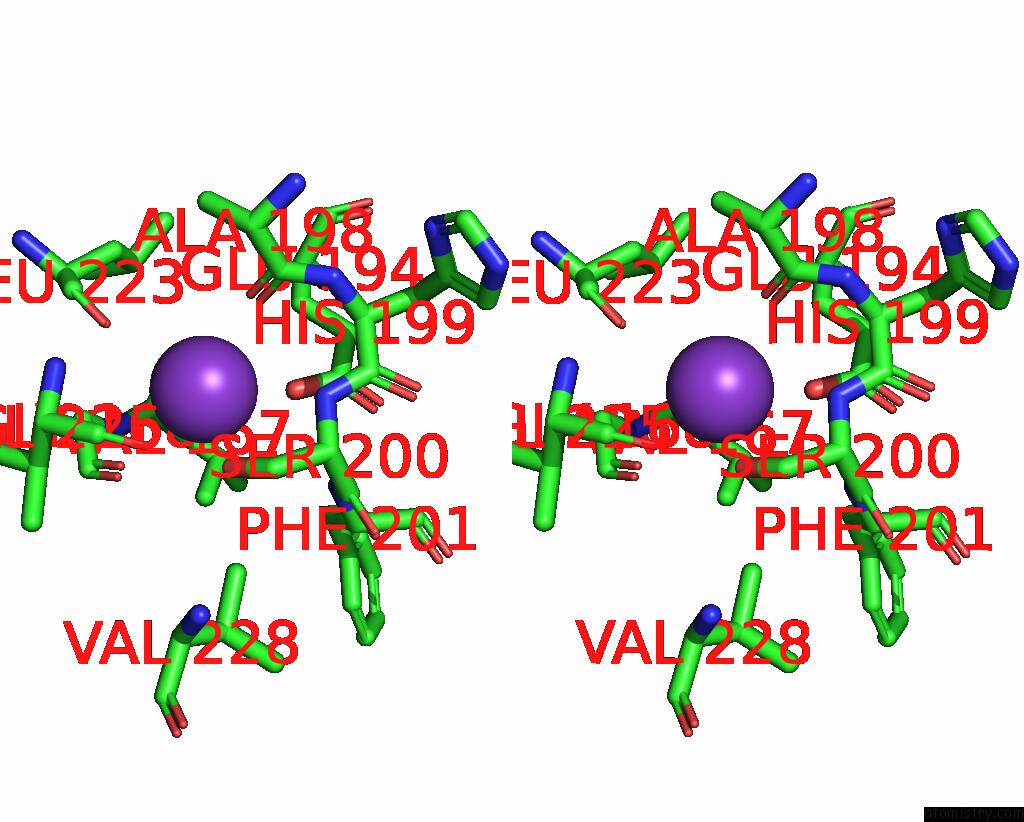
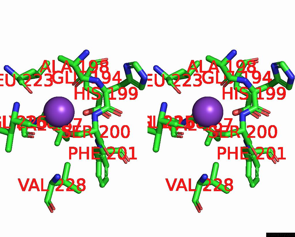
Potassium binding site 3 out of 4 in 1pwh
Go back to
Potassium binding site 3 out
of 4 in the Rat Liver L-Serine Dehydratase- Complex with Pyridoxyl-(O-Methyl- Serine)-5-Monophosphate

Mono view
Stereo pair view
Mono view
Stereo pair view
A full contact list of Potassium with other atoms in the K binding
site number 3 of Rat Liver L-Serine Dehydratase- Complex with Pyridoxyl-(O-Methyl- Serine)-5-Monophosphate within 5.0Å range:
|
Potassium binding site 4 out of 4 in 1pwh
Go back to
Potassium binding site 4 out
of 4 in the Rat Liver L-Serine Dehydratase- Complex with Pyridoxyl-(O-Methyl- Serine)-5-Monophosphate
Mono view
Stereo pair view
Mono view
Stereo pair view
A full contact list of Potassium with other atoms in the K binding
site number 4 of Rat Liver L-Serine Dehydratase- Complex with Pyridoxyl-(O-Methyl- Serine)-5-Monophosphate within 5.0Å range:
|
Reference:
T.Yamada,
J.Komoto,
Y.Takata,
H.Ogawa,
H.C.Pitot,
F.Takusagawa.
Crystal Structure of Serine Dehydratase From Rat Liver. Biochemistry V. 42 12854 2003.
ISSN: ISSN 0006-2960
PubMed: 14596599
DOI: 10.1021/BI035324P
Page generated: Sat Aug 9 02:28:55 2025
ISSN: ISSN 0006-2960
PubMed: 14596599
DOI: 10.1021/BI035324P
Last articles
Na in 4CICNa in 4CH8
Na in 4CHI
Na in 4CH2
Na in 4CG0
Na in 4CFZ
Na in 4CEY
Na in 4CFY
Na in 4CDX
Na in 4CBY