Potassium »
PDB 1o76-1pqo »
1p9e »
Potassium in PDB 1p9e: Crystal Structure Analysis of Methyl Parathion Hydrolase From Pseudomonas Sp Wbc-3
Enzymatic activity of Crystal Structure Analysis of Methyl Parathion Hydrolase From Pseudomonas Sp Wbc-3
All present enzymatic activity of Crystal Structure Analysis of Methyl Parathion Hydrolase From Pseudomonas Sp Wbc-3:
3.1.8.1;
3.1.8.1;
Protein crystallography data
The structure of Crystal Structure Analysis of Methyl Parathion Hydrolase From Pseudomonas Sp Wbc-3, PDB code: 1p9e
was solved by
Y.Dong,
L.Sun,
M.Bartlam,
Z.Rao,
X.Zhang,
with X-Ray Crystallography technique. A brief refinement statistics is given in the table below:
| Resolution Low / High (Å) | 50.00 / 2.40 |
| Space group | P 43 21 2 |
| Cell size a, b, c (Å), α, β, γ (°) | 85.020, 85.020, 199.773, 90.00, 90.00, 90.00 |
| R / Rfree (%) | 21.8 / 25.9 |
Other elements in 1p9e:
The structure of Crystal Structure Analysis of Methyl Parathion Hydrolase From Pseudomonas Sp Wbc-3 also contains other interesting chemical elements:
| Zinc | (Zn) | 3 atoms |
| Cadmium | (Cd) | 1 atom |
| Sodium | (Na) | 5 atoms |
Potassium Binding Sites:
The binding sites of Potassium atom in the Crystal Structure Analysis of Methyl Parathion Hydrolase From Pseudomonas Sp Wbc-3
(pdb code 1p9e). This binding sites where shown within
5.0 Angstroms radius around Potassium atom.
In total 4 binding sites of Potassium where determined in the Crystal Structure Analysis of Methyl Parathion Hydrolase From Pseudomonas Sp Wbc-3, PDB code: 1p9e:
Jump to Potassium binding site number: 1; 2; 3; 4;
In total 4 binding sites of Potassium where determined in the Crystal Structure Analysis of Methyl Parathion Hydrolase From Pseudomonas Sp Wbc-3, PDB code: 1p9e:
Jump to Potassium binding site number: 1; 2; 3; 4;
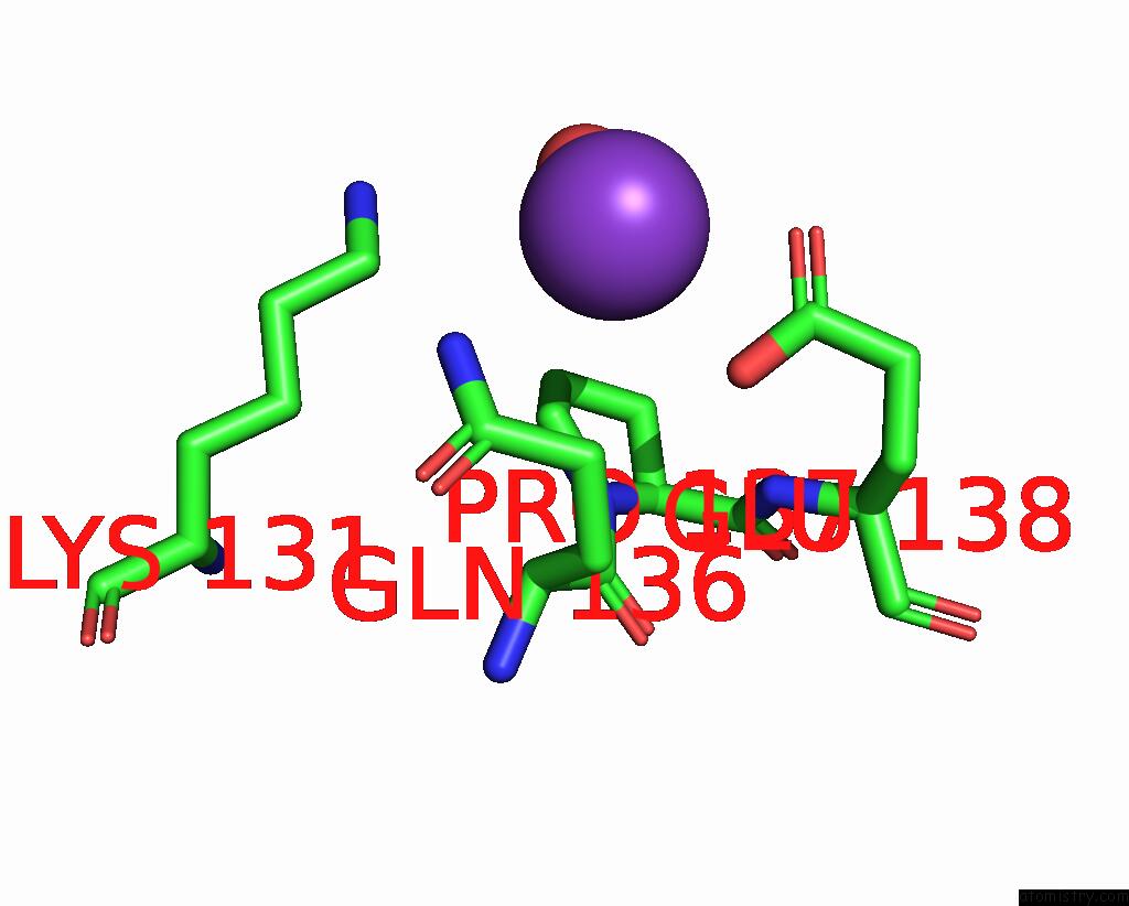
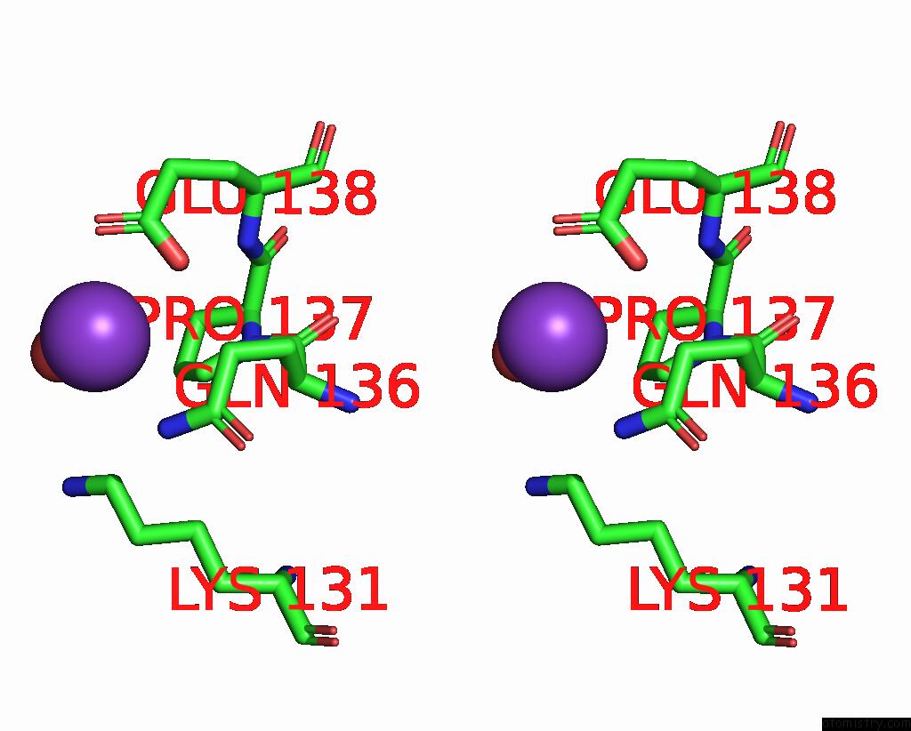
Potassium binding site 1 out of 4 in 1p9e
Go back to
Potassium binding site 1 out
of 4 in the Crystal Structure Analysis of Methyl Parathion Hydrolase From Pseudomonas Sp Wbc-3
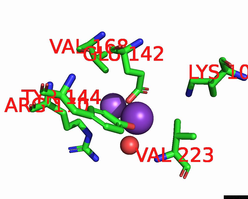
Mono view
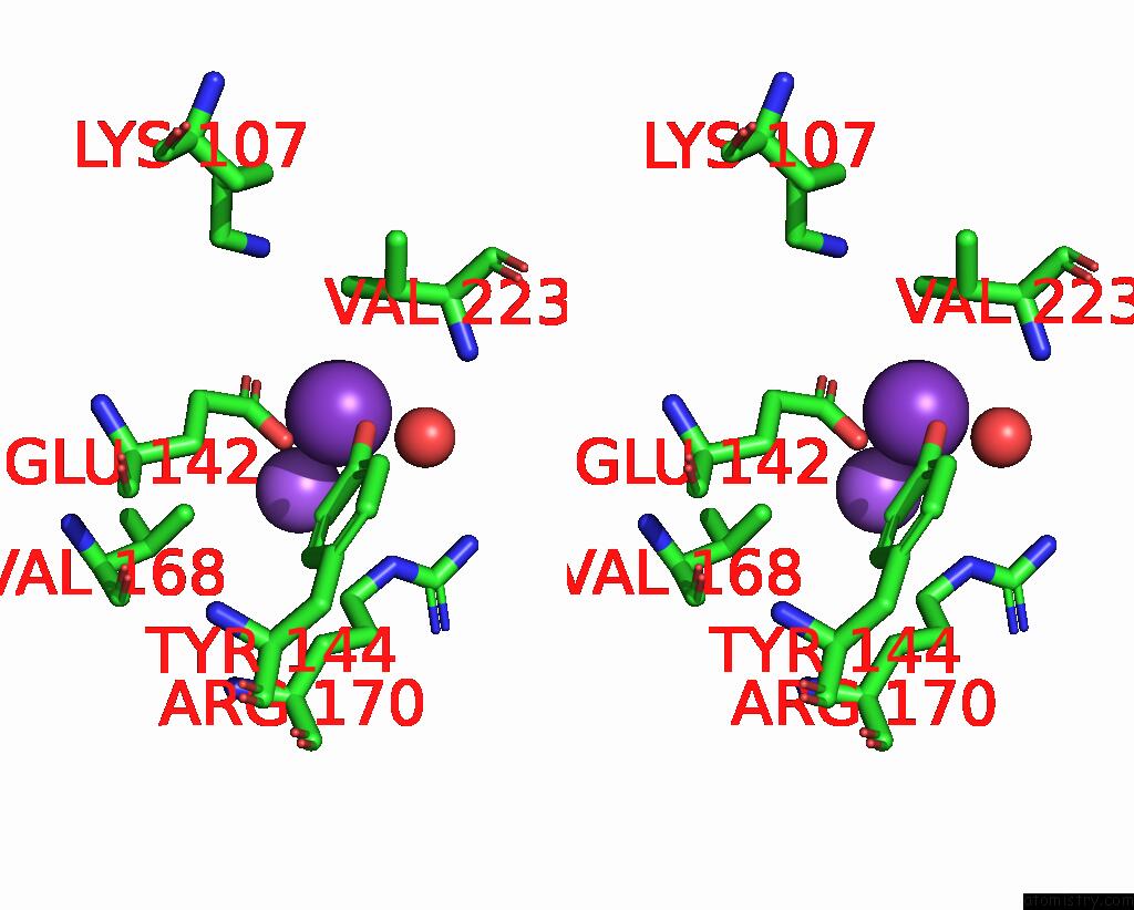
Stereo pair view
Mono view
Stereo pair view
A full contact list of Potassium with other atoms in the K binding
site number 1 of Crystal Structure Analysis of Methyl Parathion Hydrolase From Pseudomonas Sp Wbc-3 within 5.0Å range:
|
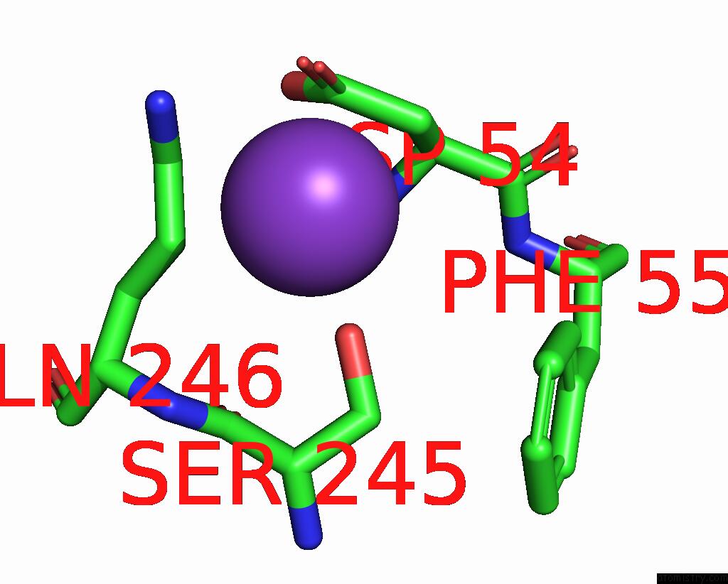
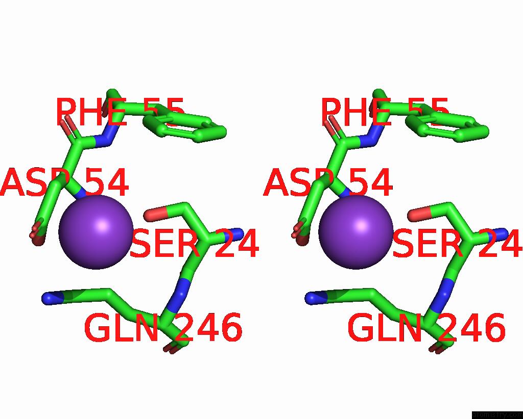
Potassium binding site 2 out of 4 in 1p9e
Go back to
Potassium binding site 2 out
of 4 in the Crystal Structure Analysis of Methyl Parathion Hydrolase From Pseudomonas Sp Wbc-3
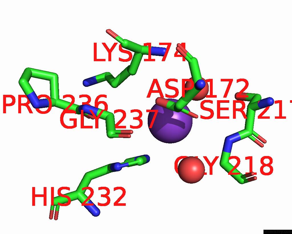
Mono view
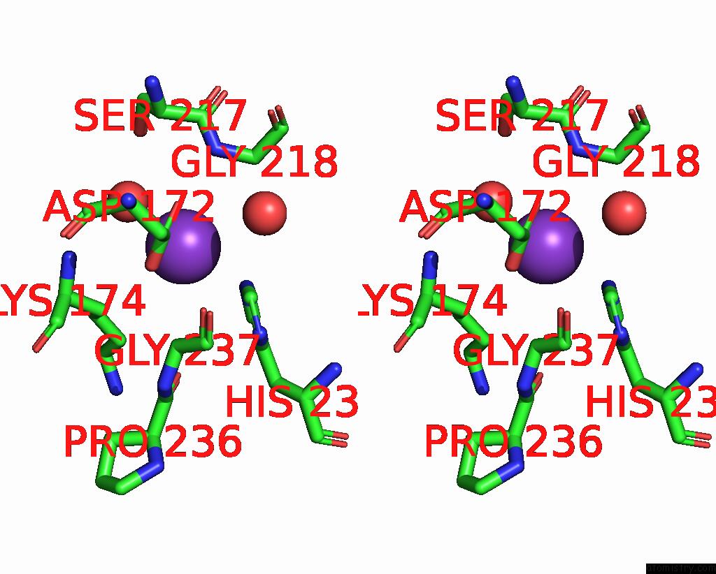
Stereo pair view
Mono view
Stereo pair view
A full contact list of Potassium with other atoms in the K binding
site number 2 of Crystal Structure Analysis of Methyl Parathion Hydrolase From Pseudomonas Sp Wbc-3 within 5.0Å range:
|
Potassium binding site 3 out of 4 in 1p9e
Go back to
Potassium binding site 3 out
of 4 in the Crystal Structure Analysis of Methyl Parathion Hydrolase From Pseudomonas Sp Wbc-3
Mono view
Stereo pair view
Mono view
Stereo pair view
A full contact list of Potassium with other atoms in the K binding
site number 3 of Crystal Structure Analysis of Methyl Parathion Hydrolase From Pseudomonas Sp Wbc-3 within 5.0Å range:
|
Potassium binding site 4 out of 4 in 1p9e
Go back to
Potassium binding site 4 out
of 4 in the Crystal Structure Analysis of Methyl Parathion Hydrolase From Pseudomonas Sp Wbc-3
Mono view
Stereo pair view
Mono view
Stereo pair view
A full contact list of Potassium with other atoms in the K binding
site number 4 of Crystal Structure Analysis of Methyl Parathion Hydrolase From Pseudomonas Sp Wbc-3 within 5.0Å range:
|
Reference:
Y.Dong,
L.Sun,
M.Bartlam,
Z.Rao,
X.Zhang.
Crystal Structure Analysis of Methyl Parathion Hydrolase From Pseudomonas Sp Wbc-3 To Be Published.
Page generated: Mon Aug 12 05:10:50 2024
Last articles
F in 8VA6F in 8VA5
F in 8V9M
F in 8V9C
F in 8V8G
F in 8V8E
F in 8V8U
F in 8V8V
F in 8V39
F in 8V7G