Potassium »
PDB 1d7v-1gmk »
1dio »
Potassium in PDB 1dio: Diol Dehydratase-Cyanocobalamin Complex From Klebsiella Oxytoca
Enzymatic activity of Diol Dehydratase-Cyanocobalamin Complex From Klebsiella Oxytoca
All present enzymatic activity of Diol Dehydratase-Cyanocobalamin Complex From Klebsiella Oxytoca:
4.2.1.28;
4.2.1.28;
Protein crystallography data
The structure of Diol Dehydratase-Cyanocobalamin Complex From Klebsiella Oxytoca, PDB code: 1dio
was solved by
N.Shibata,
J.Masuda,
T.Tobimatsu,
T.Toraya,
K.Suto,
Y.Morimoto,
N.Yasuoka,
with X-Ray Crystallography technique. A brief refinement statistics is given in the table below:
| Resolution Low / High (Å) | 10.00 / 2.20 |
| Space group | P 21 21 21 |
| Cell size a, b, c (Å), α, β, γ (°) | 76.200, 122.300, 209.600, 90.00, 90.00, 90.00 |
| R / Rfree (%) | 18.7 / 23.6 |
Other elements in 1dio:
The structure of Diol Dehydratase-Cyanocobalamin Complex From Klebsiella Oxytoca also contains other interesting chemical elements:
| Cobalt | (Co) | 2 atoms |
Potassium Binding Sites:
The binding sites of Potassium atom in the Diol Dehydratase-Cyanocobalamin Complex From Klebsiella Oxytoca
(pdb code 1dio). This binding sites where shown within
5.0 Angstroms radius around Potassium atom.
In total 2 binding sites of Potassium where determined in the Diol Dehydratase-Cyanocobalamin Complex From Klebsiella Oxytoca, PDB code: 1dio:
Jump to Potassium binding site number: 1; 2;
In total 2 binding sites of Potassium where determined in the Diol Dehydratase-Cyanocobalamin Complex From Klebsiella Oxytoca, PDB code: 1dio:
Jump to Potassium binding site number: 1; 2;
Potassium binding site 1 out of 2 in 1dio
Go back to
Potassium binding site 1 out
of 2 in the Diol Dehydratase-Cyanocobalamin Complex From Klebsiella Oxytoca
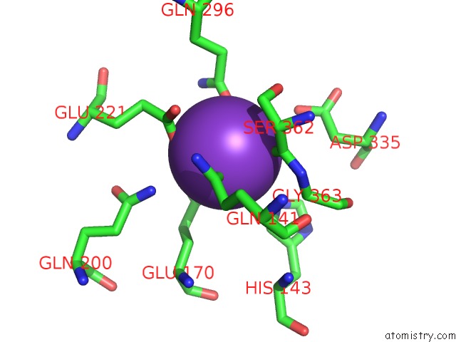
Mono view
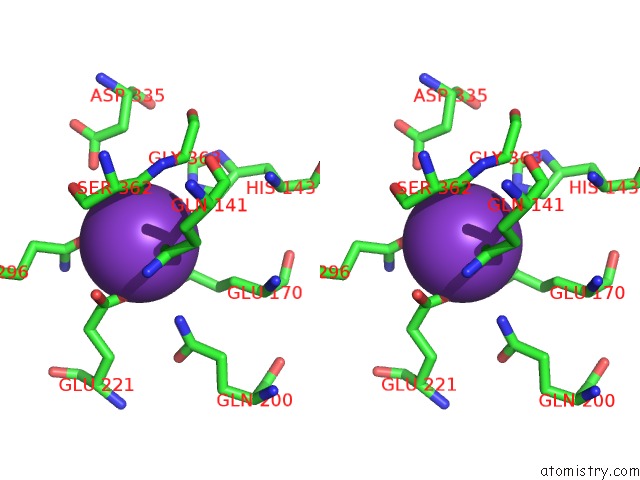
Stereo pair view
Mono view
Stereo pair view
A full contact list of Potassium with other atoms in the K binding
site number 1 of Diol Dehydratase-Cyanocobalamin Complex From Klebsiella Oxytoca within 5.0Å range:
|
Potassium binding site 2 out of 2 in 1dio
Go back to
Potassium binding site 2 out
of 2 in the Diol Dehydratase-Cyanocobalamin Complex From Klebsiella Oxytoca
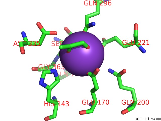
Mono view
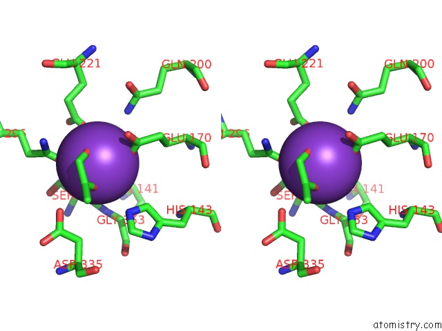
Stereo pair view
Mono view
Stereo pair view
A full contact list of Potassium with other atoms in the K binding
site number 2 of Diol Dehydratase-Cyanocobalamin Complex From Klebsiella Oxytoca within 5.0Å range:
|
Reference:
N.Shibata,
J.Masuda,
T.Tobimatsu,
T.Toraya,
K.Suto,
Y.Morimoto,
N.Yasuoka.
A New Mode of B12 Binding and the Direct Participation of A Potassium Ion in Enzyme Catalysis: X-Ray Structure of Diol Dehydratase. Structure Fold.Des. V. 7 997 1999.
ISSN: ISSN 0969-2126
PubMed: 10467140
DOI: 10.1016/S0969-2126(99)80126-9
Page generated: Sat Aug 9 01:49:54 2025
ISSN: ISSN 0969-2126
PubMed: 10467140
DOI: 10.1016/S0969-2126(99)80126-9
Last articles
W in 7ZCJW in 7RCJ
W in 7XQW
W in 7VW6
W in 7T5A
W in 7OWZ
W in 7OV7
W in 7OVS
W in 7AX2
W in 7E5Z