Potassium »
PDB 9byi-9djv »
9ccr »
Potassium in PDB 9ccr: Crystal Structure of the ESPE7 Thioesterase Mutant R35A From the Esperamicin Biosynthetic Pathway at 1.6 A
Protein crystallography data
The structure of Crystal Structure of the ESPE7 Thioesterase Mutant R35A From the Esperamicin Biosynthetic Pathway at 1.6 A, PDB code: 9ccr
was solved by
M.D.Miller,
E.D.Hankore,
W.Xu,
A.J.Kosgei,
M.Bhardwaj,
J.S.Thorson,
S.G.Vanlanen,
G.N.Phillips Jr.,
with X-Ray Crystallography technique. A brief refinement statistics is given in the table below:
| Resolution Low / High (Å) | 65.59 / 1.57 |
| Space group | P 21 21 21 |
| Cell size a, b, c (Å), α, β, γ (°) | 73.769, 90.055, 95.718, 90, 90, 90 |
| R / Rfree (%) | 17.8 / 20.4 |
Potassium Binding Sites:
The binding sites of Potassium atom in the Crystal Structure of the ESPE7 Thioesterase Mutant R35A From the Esperamicin Biosynthetic Pathway at 1.6 A
(pdb code 9ccr). This binding sites where shown within
5.0 Angstroms radius around Potassium atom.
In total 4 binding sites of Potassium where determined in the Crystal Structure of the ESPE7 Thioesterase Mutant R35A From the Esperamicin Biosynthetic Pathway at 1.6 A, PDB code: 9ccr:
Jump to Potassium binding site number: 1; 2; 3; 4;
In total 4 binding sites of Potassium where determined in the Crystal Structure of the ESPE7 Thioesterase Mutant R35A From the Esperamicin Biosynthetic Pathway at 1.6 A, PDB code: 9ccr:
Jump to Potassium binding site number: 1; 2; 3; 4;
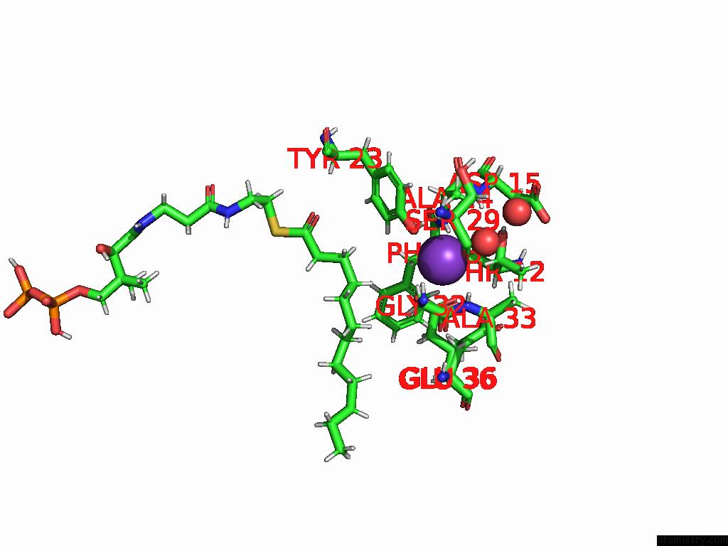
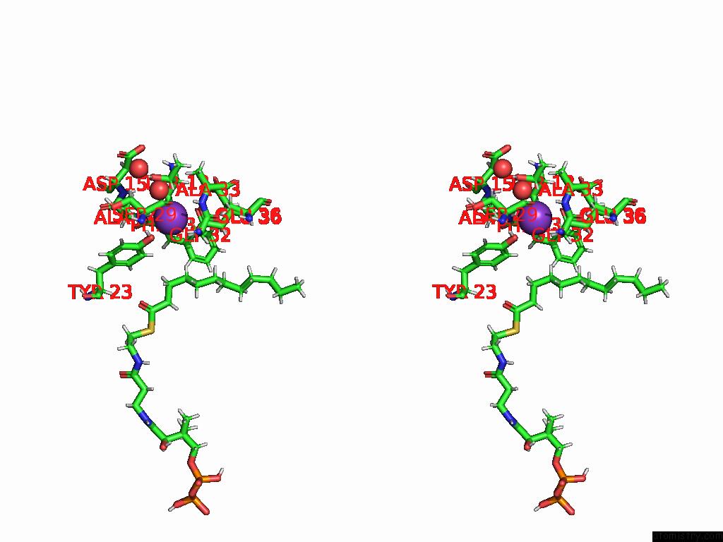
Potassium binding site 1 out of 4 in 9ccr
Go back to
Potassium binding site 1 out
of 4 in the Crystal Structure of the ESPE7 Thioesterase Mutant R35A From the Esperamicin Biosynthetic Pathway at 1.6 A
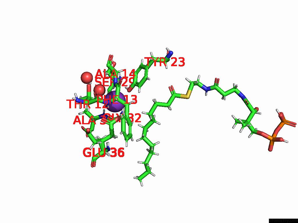
Mono view
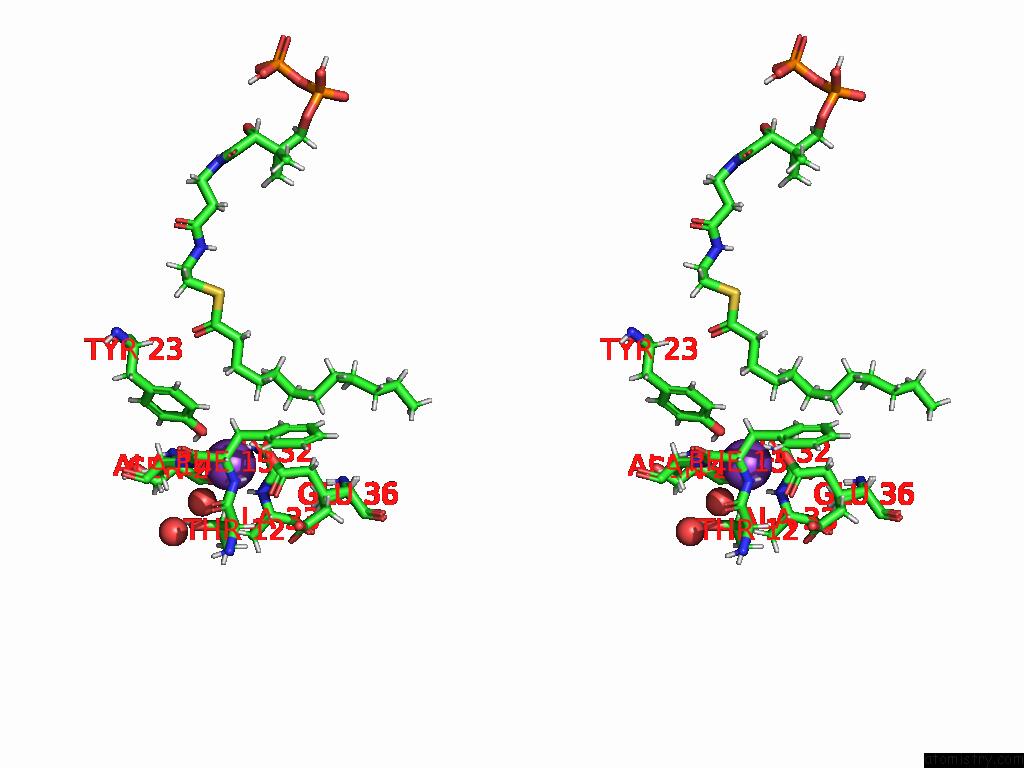
Stereo pair view
Mono view
Stereo pair view
A full contact list of Potassium with other atoms in the K binding
site number 1 of Crystal Structure of the ESPE7 Thioesterase Mutant R35A From the Esperamicin Biosynthetic Pathway at 1.6 A within 5.0Å range:
|
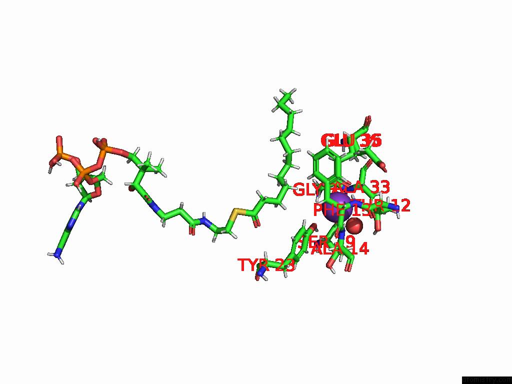
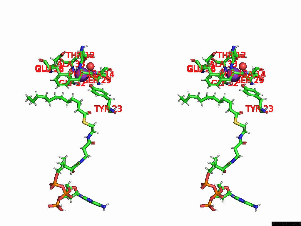
Potassium binding site 2 out of 4 in 9ccr
Go back to
Potassium binding site 2 out
of 4 in the Crystal Structure of the ESPE7 Thioesterase Mutant R35A From the Esperamicin Biosynthetic Pathway at 1.6 A
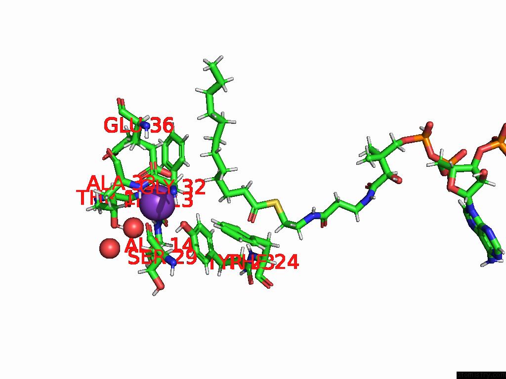
Mono view
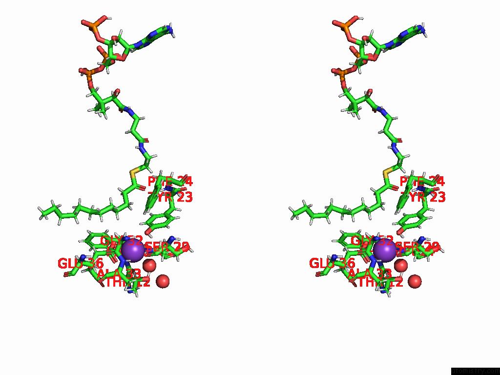
Stereo pair view
Mono view
Stereo pair view
A full contact list of Potassium with other atoms in the K binding
site number 2 of Crystal Structure of the ESPE7 Thioesterase Mutant R35A From the Esperamicin Biosynthetic Pathway at 1.6 A within 5.0Å range:
|
Potassium binding site 3 out of 4 in 9ccr
Go back to
Potassium binding site 3 out
of 4 in the Crystal Structure of the ESPE7 Thioesterase Mutant R35A From the Esperamicin Biosynthetic Pathway at 1.6 A
Mono view
Stereo pair view
Mono view
Stereo pair view
A full contact list of Potassium with other atoms in the K binding
site number 3 of Crystal Structure of the ESPE7 Thioesterase Mutant R35A From the Esperamicin Biosynthetic Pathway at 1.6 A within 5.0Å range:
|
Potassium binding site 4 out of 4 in 9ccr
Go back to
Potassium binding site 4 out
of 4 in the Crystal Structure of the ESPE7 Thioesterase Mutant R35A From the Esperamicin Biosynthetic Pathway at 1.6 A
Mono view
Stereo pair view
Mono view
Stereo pair view
A full contact list of Potassium with other atoms in the K binding
site number 4 of Crystal Structure of the ESPE7 Thioesterase Mutant R35A From the Esperamicin Biosynthetic Pathway at 1.6 A within 5.0Å range:
|
Reference:
E.D.Hankore,
M.D.Miller,
A.J.Kosgei,
W.Xu,
K.Tan,
M.Endres,
M.Bhardwaj,
G.Joachimiak,
A.Joachimiak,
G.N.Phillips Jr.,
J.S.Thorson,
S.G.Van Lanen.
Functional and Structural Studies on the Esperamicin Thioesterase and Progress Toward Understanding Enediyne Core Biosynthesis To Be Published.
Page generated: Sat Aug 9 18:34:38 2025
Last articles
Mg in 3CZJMg in 3D19
Mg in 3CZ4
Mg in 3CZ1
Mg in 3CZ0
Mg in 3CXO
Mg in 3CYZ
Mg in 3CYI
Mg in 3CX8
Mg in 3CX7