Potassium »
PDB 8sr6-8ue9 »
8u5j »
Potassium in PDB 8u5j: Structure of Mango III Variant Aptamer Bound to T01-07M-B
Protein crystallography data
The structure of Structure of Mango III Variant Aptamer Bound to T01-07M-B, PDB code: 8u5j
was solved by
L.F.M.Passalacqua,
A.R.Ferre-D'amare,
with X-Ray Crystallography technique. A brief refinement statistics is given in the table below:
| Resolution Low / High (Å) | 38.87 / 1.70 |
| Space group | P 32 2 1 |
| Cell size a, b, c (Å), α, β, γ (°) | 55.66, 55.66, 65.715, 90, 90, 120 |
| R / Rfree (%) | 20.1 / 23.6 |
Potassium Binding Sites:
The binding sites of Potassium atom in the Structure of Mango III Variant Aptamer Bound to T01-07M-B
(pdb code 8u5j). This binding sites where shown within
5.0 Angstroms radius around Potassium atom.
In total only one binding site of Potassium was determined in the Structure of Mango III Variant Aptamer Bound to T01-07M-B, PDB code: 8u5j:
In total only one binding site of Potassium was determined in the Structure of Mango III Variant Aptamer Bound to T01-07M-B, PDB code: 8u5j:
Potassium binding site 1 out of 1 in 8u5j
Go back to
Potassium binding site 1 out
of 1 in the Structure of Mango III Variant Aptamer Bound to T01-07M-B
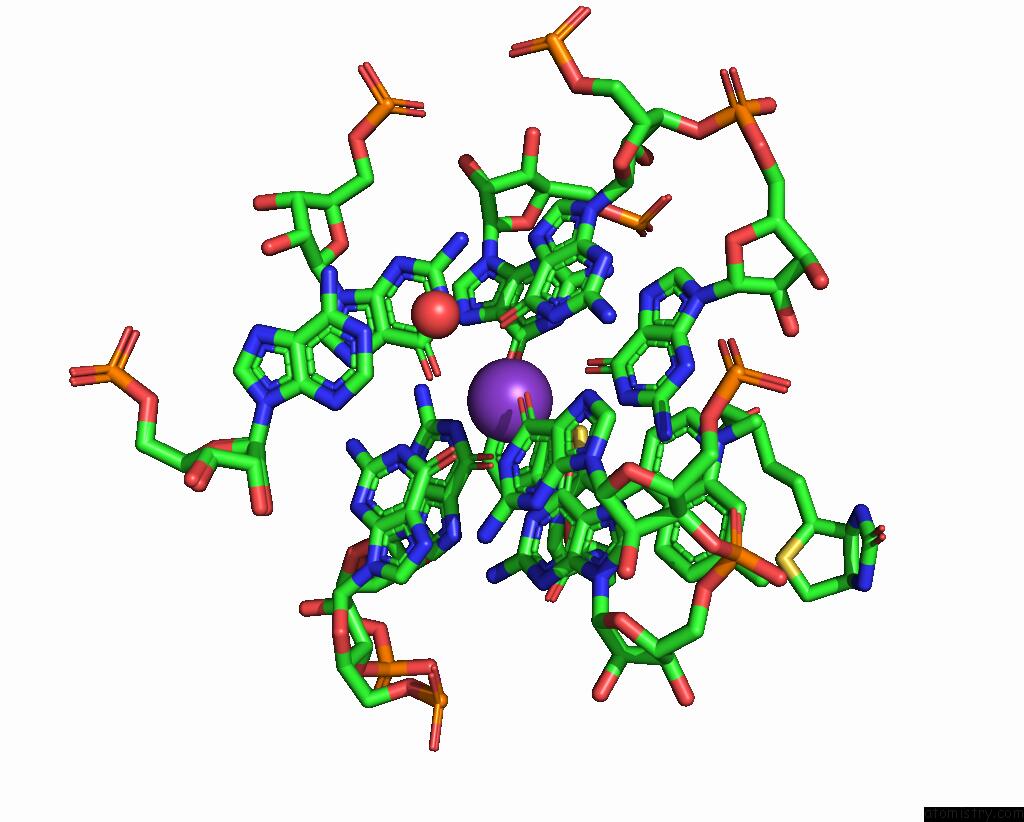
Mono view
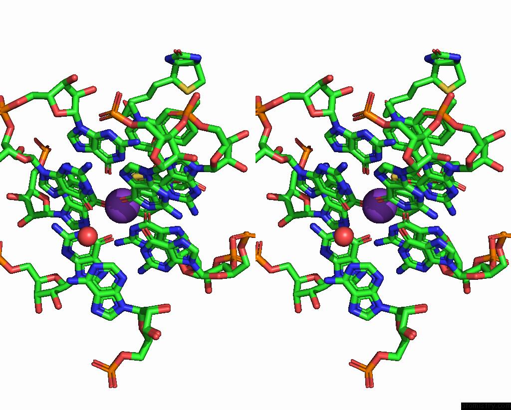
Stereo pair view
Mono view
Stereo pair view
A full contact list of Potassium with other atoms in the K binding
site number 1 of Structure of Mango III Variant Aptamer Bound to T01-07M-B within 5.0Å range:
|
Reference:
L.F.M.Passalacqua,
A.R.Ferre-D'amare.
Structure of Mango III Variant Aptamer Bound to T01-07M-B To Be Published.
Page generated: Sat Aug 9 17:57:01 2025
Last articles
Mg in 2HNYMg in 2HMF
Mg in 2HMC
Mg in 2HND
Mg in 2HMA
Mg in 2HJ6
Mg in 2HK6
Mg in 2HKJ
Mg in 2HIT
Mg in 2HJP