Potassium »
PDB 8sr6-8ue9 »
8u3n »
Potassium in PDB 8u3n: Structure of P450BLT From Micromonospora Sp. Mw-13
Protein crystallography data
The structure of Structure of P450BLT From Micromonospora Sp. Mw-13, PDB code: 8u3n
was solved by
M.H.Hansen,
M.J.Cryle,
Y.Zhao,
with X-Ray Crystallography technique. A brief refinement statistics is given in the table below:
| Resolution Low / High (Å) | 47.87 / 2.15 |
| Space group | P 1 21 1 |
| Cell size a, b, c (Å), α, β, γ (°) | 61.078, 95.748, 105.217, 90, 92.63, 90 |
| R / Rfree (%) | 19.4 / 23.6 |
Other elements in 8u3n:
The structure of Structure of P450BLT From Micromonospora Sp. Mw-13 also contains other interesting chemical elements:
| Iron | (Fe) | 3 atoms |
Potassium Binding Sites:
The binding sites of Potassium atom in the Structure of P450BLT From Micromonospora Sp. Mw-13
(pdb code 8u3n). This binding sites where shown within
5.0 Angstroms radius around Potassium atom.
In total 3 binding sites of Potassium where determined in the Structure of P450BLT From Micromonospora Sp. Mw-13, PDB code: 8u3n:
Jump to Potassium binding site number: 1; 2; 3;
In total 3 binding sites of Potassium where determined in the Structure of P450BLT From Micromonospora Sp. Mw-13, PDB code: 8u3n:
Jump to Potassium binding site number: 1; 2; 3;
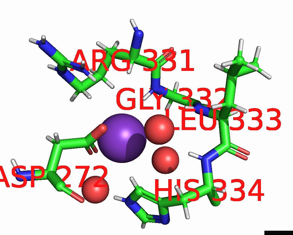
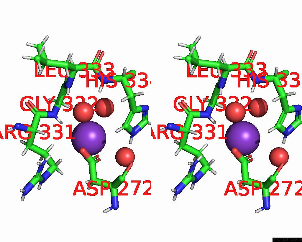
Potassium binding site 1 out of 3 in 8u3n
Go back to
Potassium binding site 1 out
of 3 in the Structure of P450BLT From Micromonospora Sp. Mw-13
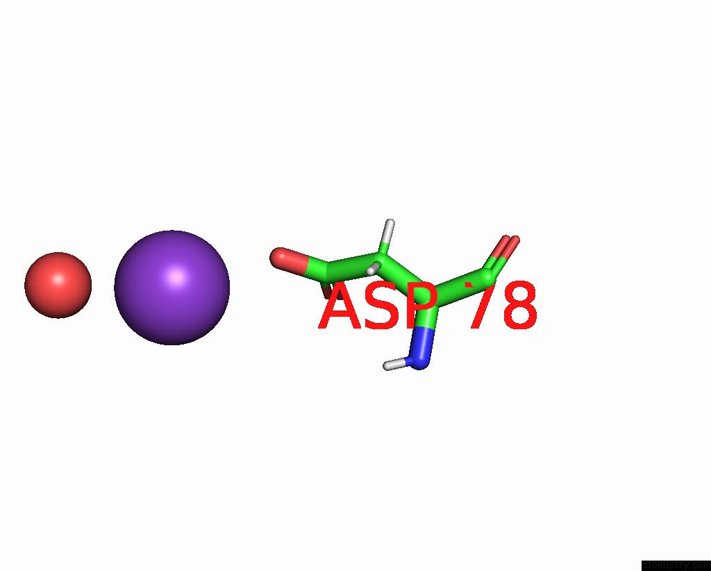
Mono view
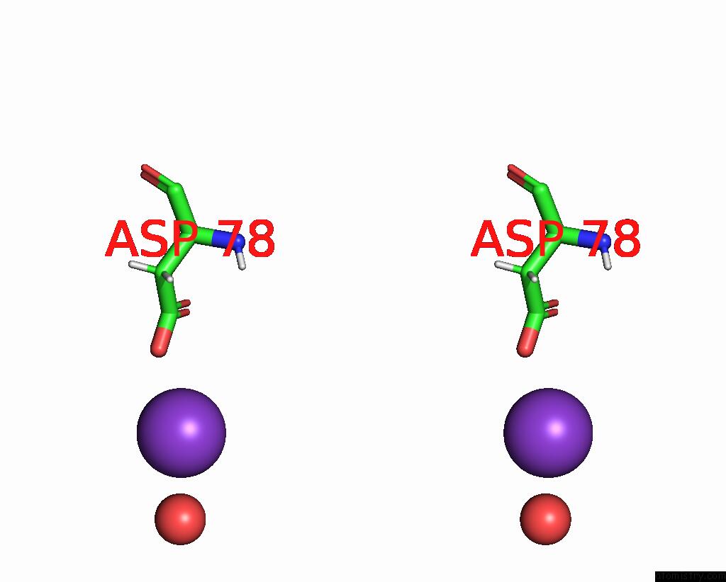
Stereo pair view
Mono view
Stereo pair view
A full contact list of Potassium with other atoms in the K binding
site number 1 of Structure of P450BLT From Micromonospora Sp. Mw-13 within 5.0Å range:
|
Potassium binding site 2 out of 3 in 8u3n
Go back to
Potassium binding site 2 out
of 3 in the Structure of P450BLT From Micromonospora Sp. Mw-13
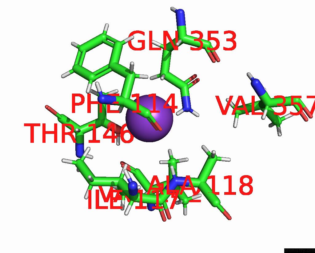
Mono view
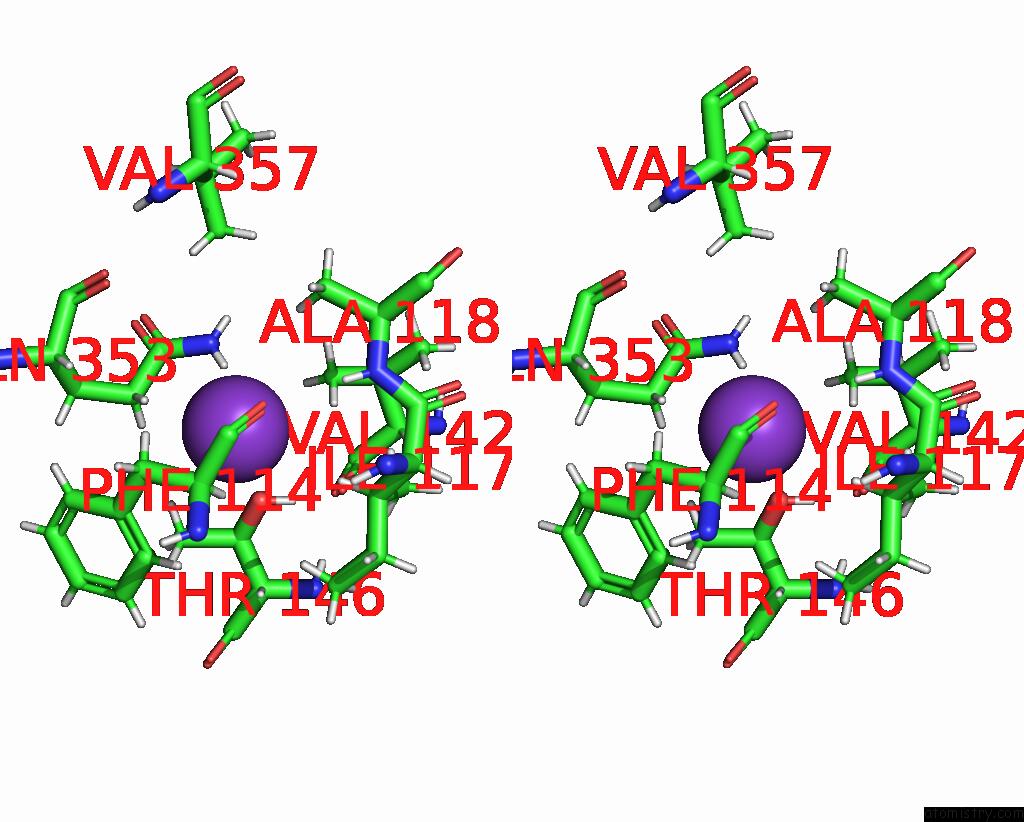
Stereo pair view
Mono view
Stereo pair view
A full contact list of Potassium with other atoms in the K binding
site number 2 of Structure of P450BLT From Micromonospora Sp. Mw-13 within 5.0Å range:
|
Potassium binding site 3 out of 3 in 8u3n
Go back to
Potassium binding site 3 out
of 3 in the Structure of P450BLT From Micromonospora Sp. Mw-13
Mono view
Stereo pair view
Mono view
Stereo pair view
A full contact list of Potassium with other atoms in the K binding
site number 3 of Structure of P450BLT From Micromonospora Sp. Mw-13 within 5.0Å range:
|
Reference:
M.H.Hansen,
A.Keto,
M.Treisman,
V.M.Sasi,
L.Coe,
Y.Zhao,
L.Padva,
C.Hess,
V.Leichthammer,
D.L.Machell,
R.B.Schittenhelm,
C.J.Jackson,
J.Tailhades,
M.Crusemann,
J.J.De Voss,
E.H.Krenske,
M.J.Cryle.
Structural Insights Into A Side Chain Cross-Linking Biarylitide P450 From Ripp Biosynthesis Acs Catalysis 812 2024.
ISSN: ESSN 2155-5435
DOI: 10.1021/ACSCATAL.3C05417
Page generated: Sat Aug 9 17:56:49 2025
ISSN: ESSN 2155-5435
DOI: 10.1021/ACSCATAL.3C05417
Last articles
Mg in 1FIRMg in 1FI1
Mg in 1FHV
Mg in 1FGS
Mg in 1FEZ
Mg in 1FFK
Mg in 1FFH
Mg in 1FC5
Mg in 1FDG
Mg in 1FD5