Potassium »
PDB 8qrk-8sfz »
8s1x »
Potassium in PDB 8s1x: Crystal Structure of Actinonin-Bound PDF1 and the Computationally Designed DBACT553_1 Protein Binder
Enzymatic activity of Crystal Structure of Actinonin-Bound PDF1 and the Computationally Designed DBACT553_1 Protein Binder
All present enzymatic activity of Crystal Structure of Actinonin-Bound PDF1 and the Computationally Designed DBACT553_1 Protein Binder:
3.5.1.88;
3.5.1.88;
Protein crystallography data
The structure of Crystal Structure of Actinonin-Bound PDF1 and the Computationally Designed DBACT553_1 Protein Binder, PDB code: 8s1x
was solved by
A.Marchand,
M.Pacesa,
B.E.Correia,
with X-Ray Crystallography technique. A brief refinement statistics is given in the table below:
| Resolution Low / High (Å) | 55.70 / 1.88 |
| Space group | P 21 21 21 |
| Cell size a, b, c (Å), α, β, γ (°) | 49.44, 75.01, 83.16, 90, 90, 90 |
| R / Rfree (%) | 18.4 / 20.3 |
Other elements in 8s1x:
The structure of Crystal Structure of Actinonin-Bound PDF1 and the Computationally Designed DBACT553_1 Protein Binder also contains other interesting chemical elements:
| Zinc | (Zn) | 1 atom |
Potassium Binding Sites:
The binding sites of Potassium atom in the Crystal Structure of Actinonin-Bound PDF1 and the Computationally Designed DBACT553_1 Protein Binder
(pdb code 8s1x). This binding sites where shown within
5.0 Angstroms radius around Potassium atom.
In total only one binding site of Potassium was determined in the Crystal Structure of Actinonin-Bound PDF1 and the Computationally Designed DBACT553_1 Protein Binder, PDB code: 8s1x:
In total only one binding site of Potassium was determined in the Crystal Structure of Actinonin-Bound PDF1 and the Computationally Designed DBACT553_1 Protein Binder, PDB code: 8s1x:
Potassium binding site 1 out of 1 in 8s1x
Go back to
Potassium binding site 1 out
of 1 in the Crystal Structure of Actinonin-Bound PDF1 and the Computationally Designed DBACT553_1 Protein Binder
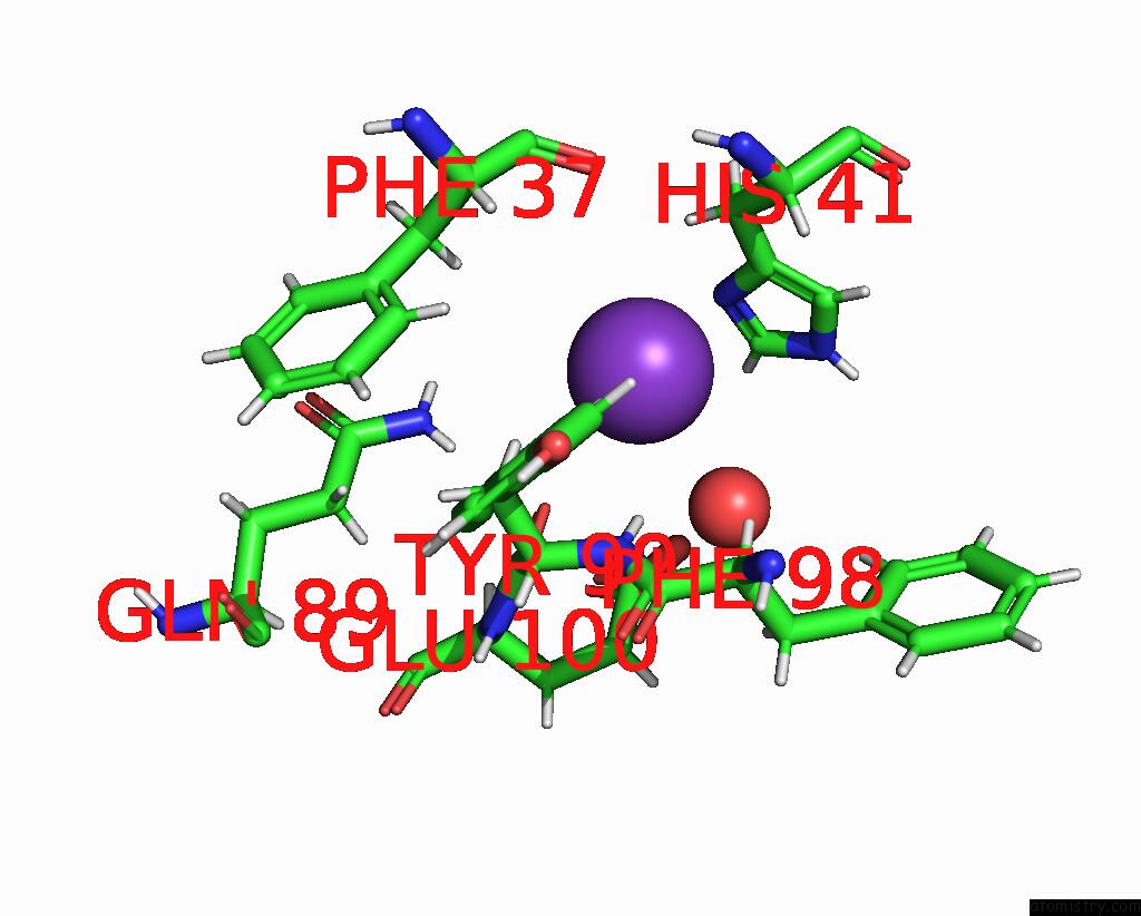
Mono view
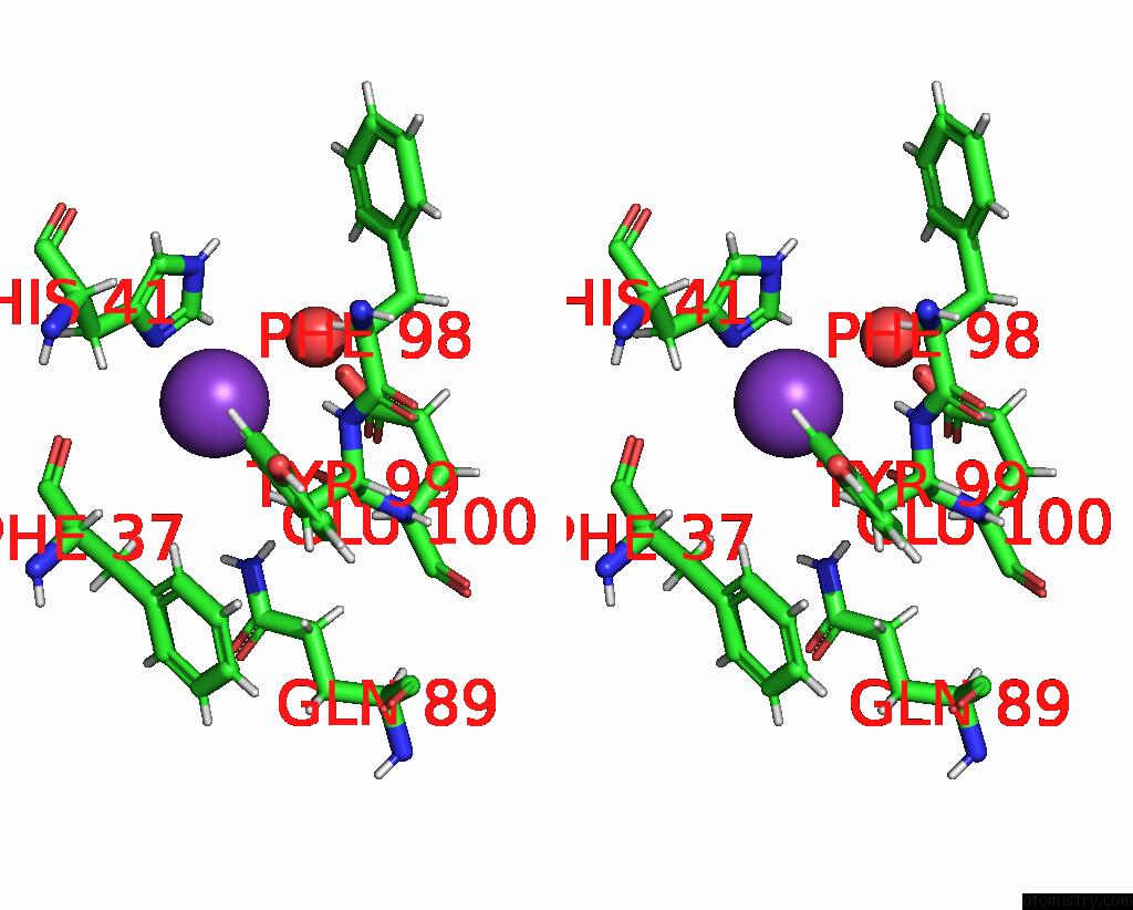
Stereo pair view
Mono view
Stereo pair view
A full contact list of Potassium with other atoms in the K binding
site number 1 of Crystal Structure of Actinonin-Bound PDF1 and the Computationally Designed DBACT553_1 Protein Binder within 5.0Å range:
|
Reference:
A.Marchand,
S.Buckley,
A.Schneuing,
M.Pacesa,
P.Gainza,
E.Elizarova,
R.Neeser,
L.Reymond,
S.Georgeon,
J.Schmidt,
B.E.Correia.
Targeting Hybrid Neosurface Fingerprints For the Design of De Novo Drug-Induced Protein Interactions To Be Published.
Page generated: Sat Aug 9 17:46:08 2025
Last articles
Mg in 2X9FMg in 2X9H
Mg in 2X7D
Mg in 2X7E
Mg in 2X7C
Mg in 2X6S
Mg in 2X6U
Mg in 2X7A
Mg in 2X6V
Mg in 2X77