Potassium »
PDB 8ooi-8qos »
8qh9 »
Potassium in PDB 8qh9: X-Ray Structure of Danio Rerio Histone Deacetylase 6 (HDAC6) CD2 in Complex with A S-29B
Protein crystallography data
The structure of X-Ray Structure of Danio Rerio Histone Deacetylase 6 (HDAC6) CD2 in Complex with A S-29B, PDB code: 8qh9
was solved by
C.Barinka,
L.Motlova,
with X-Ray Crystallography technique. A brief refinement statistics is given in the table below:
| Resolution Low / High (Å) | 47.13 / 1.59 |
| Space group | P 21 21 2 |
| Cell size a, b, c (Å), α, β, γ (°) | 84.39, 94.26, 51.65, 90, 90, 90 |
| R / Rfree (%) | 19.7 / 22.6 |
Other elements in 8qh9:
The structure of X-Ray Structure of Danio Rerio Histone Deacetylase 6 (HDAC6) CD2 in Complex with A S-29B also contains other interesting chemical elements:
| Zinc | (Zn) | 1 atom |
Potassium Binding Sites:
The binding sites of Potassium atom in the X-Ray Structure of Danio Rerio Histone Deacetylase 6 (HDAC6) CD2 in Complex with A S-29B
(pdb code 8qh9). This binding sites where shown within
5.0 Angstroms radius around Potassium atom.
In total 2 binding sites of Potassium where determined in the X-Ray Structure of Danio Rerio Histone Deacetylase 6 (HDAC6) CD2 in Complex with A S-29B, PDB code: 8qh9:
Jump to Potassium binding site number: 1; 2;
In total 2 binding sites of Potassium where determined in the X-Ray Structure of Danio Rerio Histone Deacetylase 6 (HDAC6) CD2 in Complex with A S-29B, PDB code: 8qh9:
Jump to Potassium binding site number: 1; 2;
Potassium binding site 1 out of 2 in 8qh9
Go back to
Potassium binding site 1 out
of 2 in the X-Ray Structure of Danio Rerio Histone Deacetylase 6 (HDAC6) CD2 in Complex with A S-29B
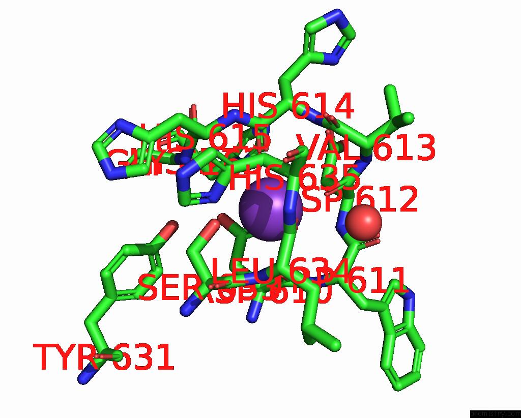
Mono view
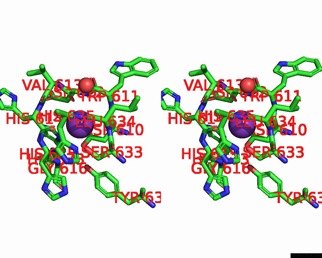
Stereo pair view
Mono view
Stereo pair view
A full contact list of Potassium with other atoms in the K binding
site number 1 of X-Ray Structure of Danio Rerio Histone Deacetylase 6 (HDAC6) CD2 in Complex with A S-29B within 5.0Å range:
|
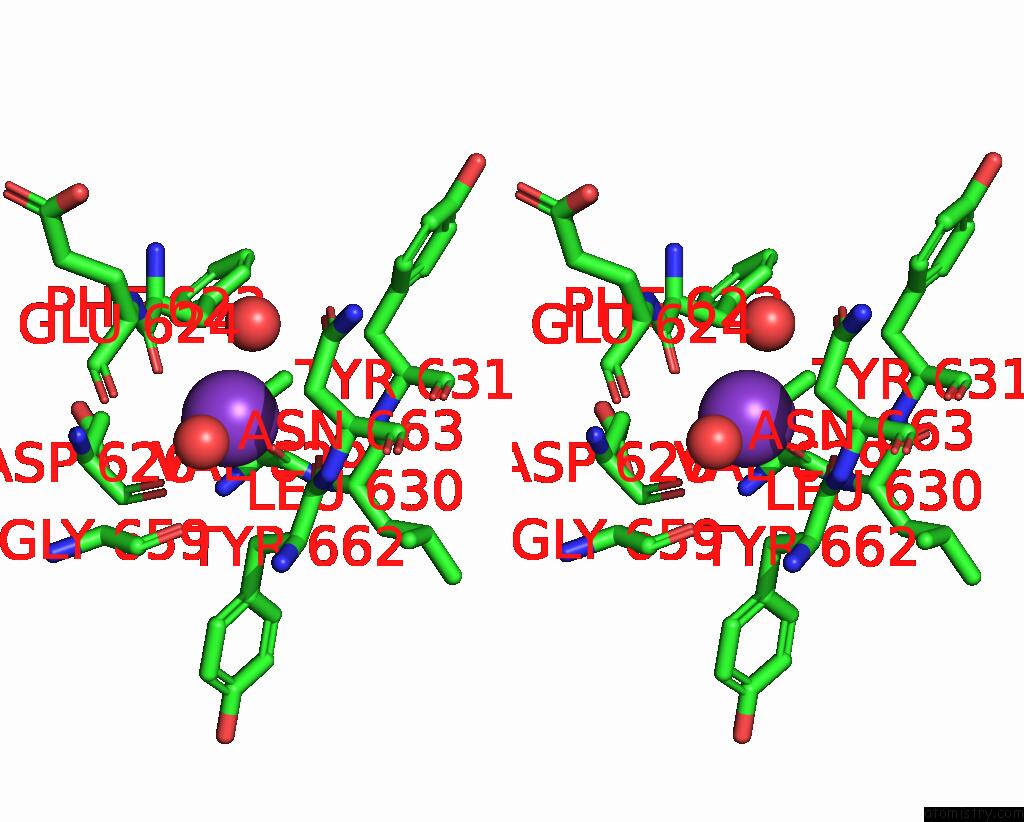
Potassium binding site 2 out of 2 in 8qh9
Go back to
Potassium binding site 2 out
of 2 in the X-Ray Structure of Danio Rerio Histone Deacetylase 6 (HDAC6) CD2 in Complex with A S-29B

Mono view
Stereo pair view
Mono view
Stereo pair view
A full contact list of Potassium with other atoms in the K binding
site number 2 of X-Ray Structure of Danio Rerio Histone Deacetylase 6 (HDAC6) CD2 in Complex with A S-29B within 5.0Å range:
|
Reference:
S.Scheuerer,
L.Motlova,
L.Schaker-Hubner,
A.Sellmer,
F.Feller,
F.J.Ertl,
P.Koch,
F.K.Hansen,
C.Barinka,
S.Mahboobi.
Biological and Structural Investigation of Tetrahydro-Beta-Carboline-Based Selective HDAC6 Inhibitors with Improved Stability. Eur.J.Med.Chem. V. 276 16676 2024.
ISSN: ISSN 0223-5234
PubMed: 39067437
DOI: 10.1016/J.EJMECH.2024.116676
Page generated: Sat Aug 9 17:28:51 2025
ISSN: ISSN 0223-5234
PubMed: 39067437
DOI: 10.1016/J.EJMECH.2024.116676
Last articles
Mg in 3WAGMg in 3W7F
Mg in 3WAD
Mg in 3W9S
Mg in 3W7T
Mg in 3W6S
Mg in 3W6P
Mg in 3W6O
Mg in 3W5A
Mg in 3W6N