Potassium »
PDB 8ooi-8qos »
8qa7 »
Potassium in PDB 8qa7: Crystal Structure of HDAC6 Catalytic Domain 2 From Zebrafish in Complex with Buffer Component.
Protein crystallography data
The structure of Crystal Structure of HDAC6 Catalytic Domain 2 From Zebrafish in Complex with Buffer Component., PDB code: 8qa7
was solved by
J.Sandmark,
M.Ek,
with X-Ray Crystallography technique. A brief refinement statistics is given in the table below:
| Resolution Low / High (Å) | 66.63 / 1.47 |
| Space group | P 21 21 21 |
| Cell size a, b, c (Å), α, β, γ (°) | 75.39, 92.373, 96.19, 90, 90, 90 |
| R / Rfree (%) | 21.7 / 24 |
Other elements in 8qa7:
The structure of Crystal Structure of HDAC6 Catalytic Domain 2 From Zebrafish in Complex with Buffer Component. also contains other interesting chemical elements:
| Zinc | (Zn) | 2 atoms |
Potassium Binding Sites:
The binding sites of Potassium atom in the Crystal Structure of HDAC6 Catalytic Domain 2 From Zebrafish in Complex with Buffer Component.
(pdb code 8qa7). This binding sites where shown within
5.0 Angstroms radius around Potassium atom.
In total 4 binding sites of Potassium where determined in the Crystal Structure of HDAC6 Catalytic Domain 2 From Zebrafish in Complex with Buffer Component., PDB code: 8qa7:
Jump to Potassium binding site number: 1; 2; 3; 4;
In total 4 binding sites of Potassium where determined in the Crystal Structure of HDAC6 Catalytic Domain 2 From Zebrafish in Complex with Buffer Component., PDB code: 8qa7:
Jump to Potassium binding site number: 1; 2; 3; 4;
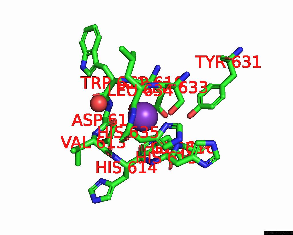
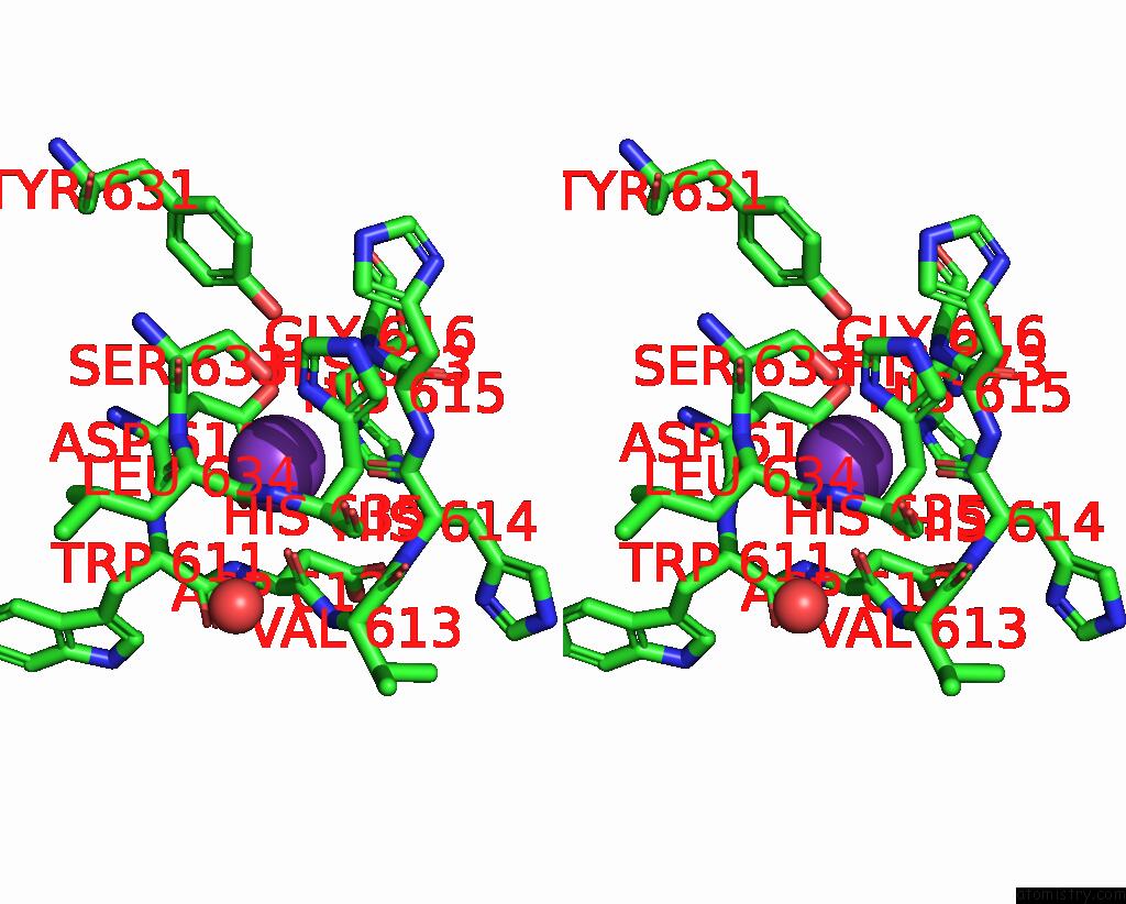
Potassium binding site 1 out of 4 in 8qa7
Go back to
Potassium binding site 1 out
of 4 in the Crystal Structure of HDAC6 Catalytic Domain 2 From Zebrafish in Complex with Buffer Component.
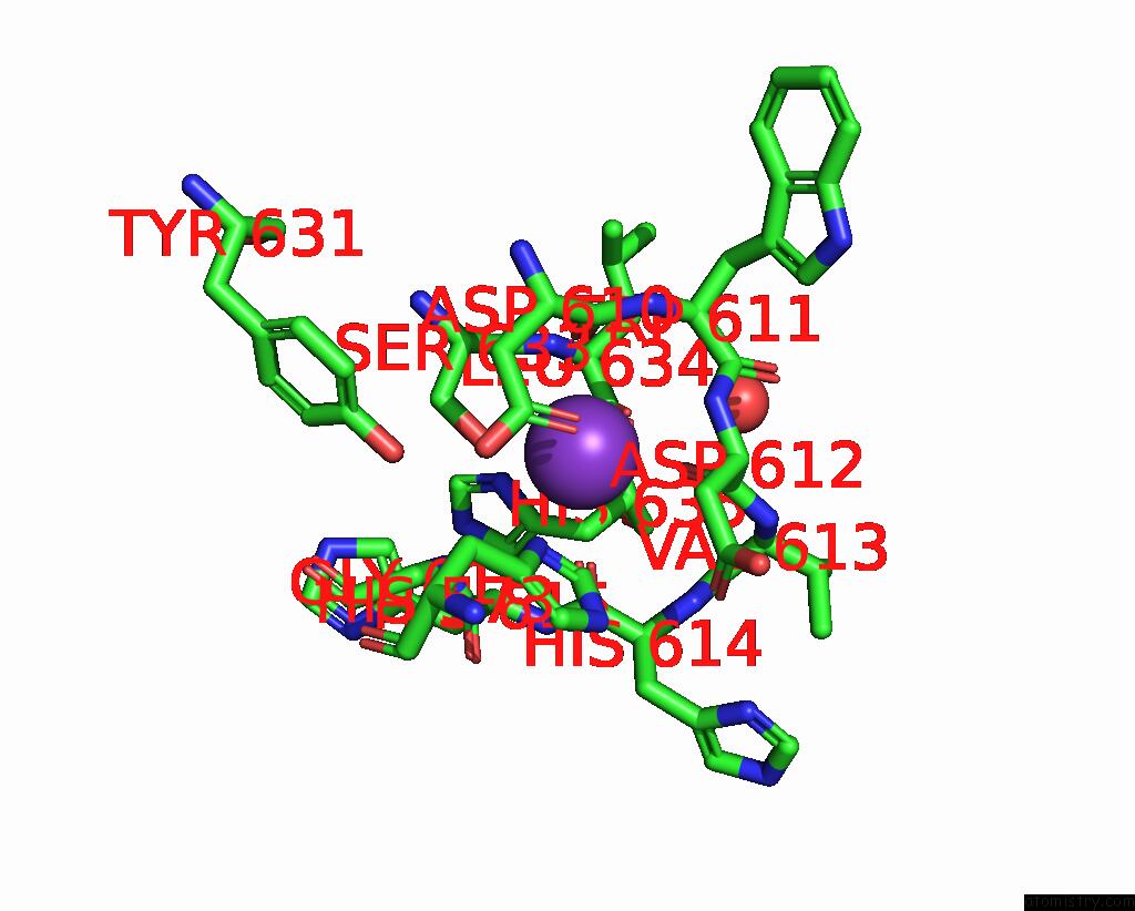
Mono view
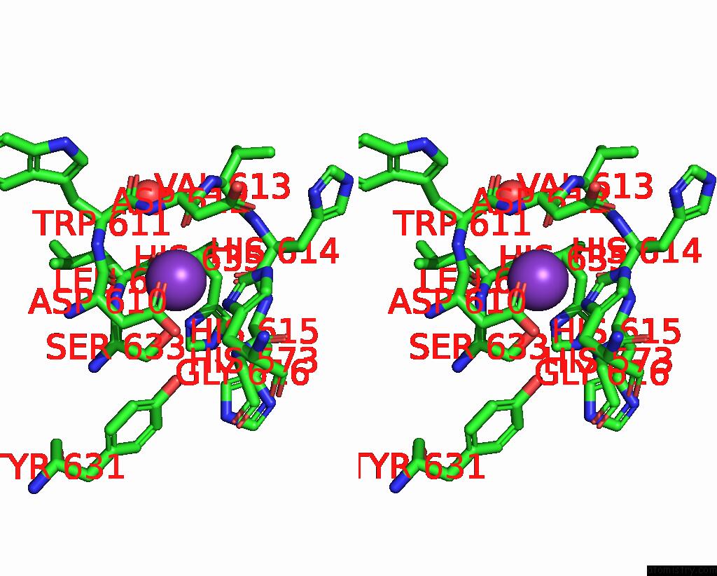
Stereo pair view
Mono view
Stereo pair view
A full contact list of Potassium with other atoms in the K binding
site number 1 of Crystal Structure of HDAC6 Catalytic Domain 2 From Zebrafish in Complex with Buffer Component. within 5.0Å range:
|
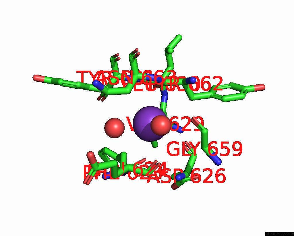
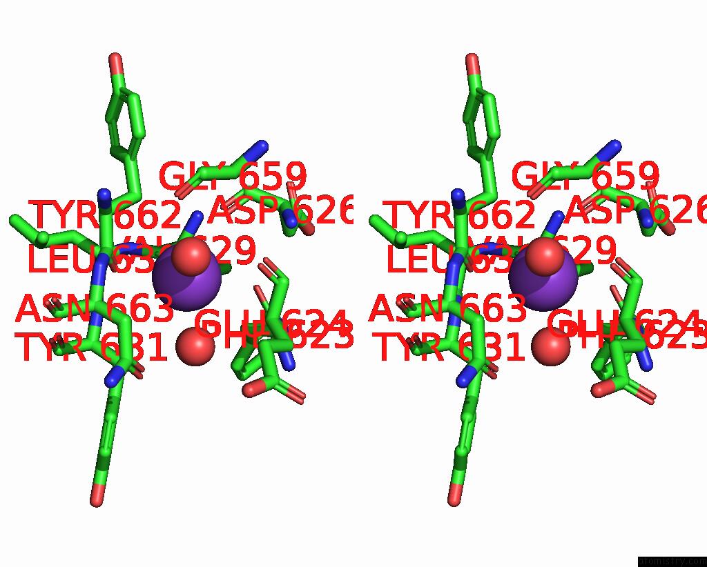
Potassium binding site 2 out of 4 in 8qa7
Go back to
Potassium binding site 2 out
of 4 in the Crystal Structure of HDAC6 Catalytic Domain 2 From Zebrafish in Complex with Buffer Component.
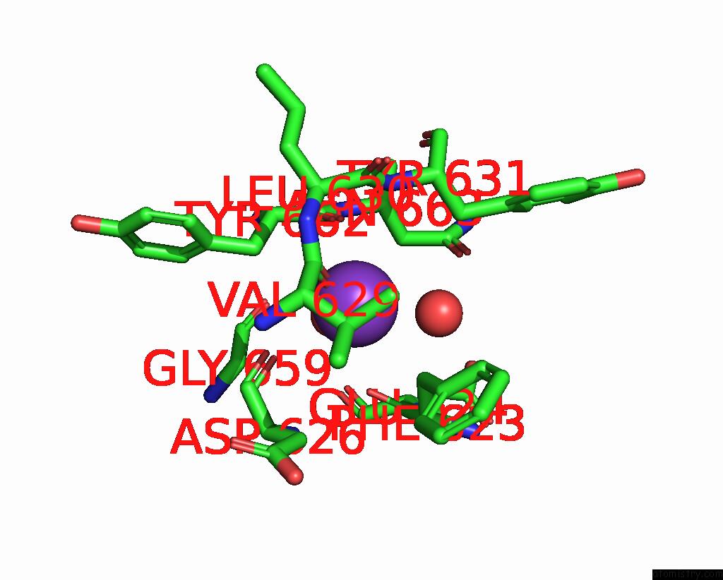
Mono view
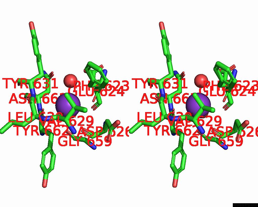
Stereo pair view
Mono view
Stereo pair view
A full contact list of Potassium with other atoms in the K binding
site number 2 of Crystal Structure of HDAC6 Catalytic Domain 2 From Zebrafish in Complex with Buffer Component. within 5.0Å range:
|
Potassium binding site 3 out of 4 in 8qa7
Go back to
Potassium binding site 3 out
of 4 in the Crystal Structure of HDAC6 Catalytic Domain 2 From Zebrafish in Complex with Buffer Component.
Mono view
Stereo pair view
Mono view
Stereo pair view
A full contact list of Potassium with other atoms in the K binding
site number 3 of Crystal Structure of HDAC6 Catalytic Domain 2 From Zebrafish in Complex with Buffer Component. within 5.0Å range:
|
Potassium binding site 4 out of 4 in 8qa7
Go back to
Potassium binding site 4 out
of 4 in the Crystal Structure of HDAC6 Catalytic Domain 2 From Zebrafish in Complex with Buffer Component.
Mono view
Stereo pair view
Mono view
Stereo pair view
A full contact list of Potassium with other atoms in the K binding
site number 4 of Crystal Structure of HDAC6 Catalytic Domain 2 From Zebrafish in Complex with Buffer Component. within 5.0Å range:
|
Reference:
J.Sandmark,
M.Ek.
Crystal Structure of HDAC6 Catalytic Domain 2 From Zebrafish in Complex with Buffer Component. To Be Published.
Page generated: Sat Aug 9 17:28:30 2025
Last articles
Mg in 3VYSMg in 3VVH
Mg in 3VX4
Mg in 3VV1
Mg in 3VTI
Mg in 3VWK
Mg in 3VWJ
Mg in 3VTH
Mg in 3VR6
Mg in 3VR3