Potassium »
PDB 8hzf-8olz »
8ipp »
Potassium in PDB 8ipp: Crystal Structure of the Complex Between An Ankyrin and A Parallel G- Quadruplex
Protein crystallography data
The structure of Crystal Structure of the Complex Between An Ankyrin and A Parallel G- Quadruplex, PDB code: 8ipp
was solved by
K.H.Ngo,
C.W.Liew,
B.Heddi,
A.T.Phan,
with X-Ray Crystallography technique. A brief refinement statistics is given in the table below:
| Resolution Low / High (Å) | 38.38 / 2.01 |
| Space group | P 21 21 2 |
| Cell size a, b, c (Å), α, β, γ (°) | 76.759, 83.587, 31.858, 90, 90, 90 |
| R / Rfree (%) | 21.1 / 22.4 |
Other elements in 8ipp:
The structure of Crystal Structure of the Complex Between An Ankyrin and A Parallel G- Quadruplex also contains other interesting chemical elements:
| Magnesium | (Mg) | 2 atoms |
Potassium Binding Sites:
The binding sites of Potassium atom in the Crystal Structure of the Complex Between An Ankyrin and A Parallel G- Quadruplex
(pdb code 8ipp). This binding sites where shown within
5.0 Angstroms radius around Potassium atom.
In total 3 binding sites of Potassium where determined in the Crystal Structure of the Complex Between An Ankyrin and A Parallel G- Quadruplex, PDB code: 8ipp:
Jump to Potassium binding site number: 1; 2; 3;
In total 3 binding sites of Potassium where determined in the Crystal Structure of the Complex Between An Ankyrin and A Parallel G- Quadruplex, PDB code: 8ipp:
Jump to Potassium binding site number: 1; 2; 3;
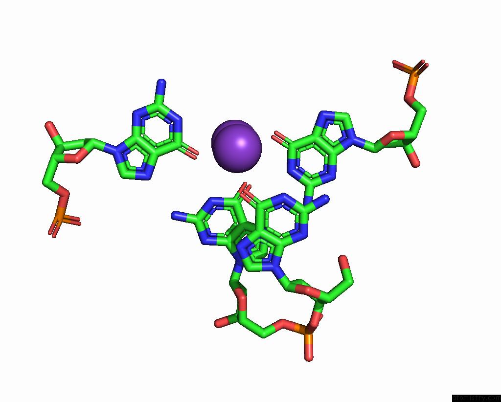
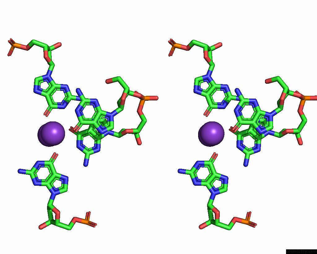
Potassium binding site 1 out of 3 in 8ipp
Go back to
Potassium binding site 1 out
of 3 in the Crystal Structure of the Complex Between An Ankyrin and A Parallel G- Quadruplex
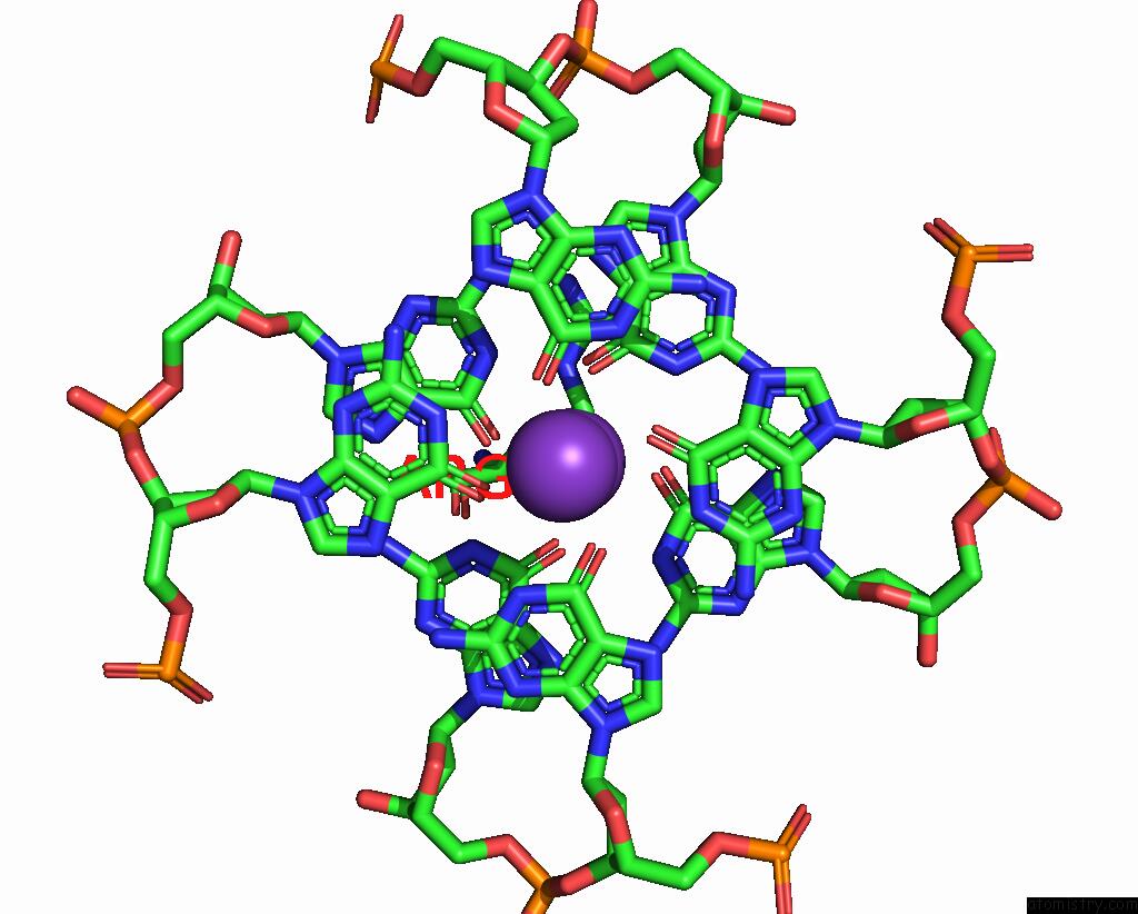
Mono view
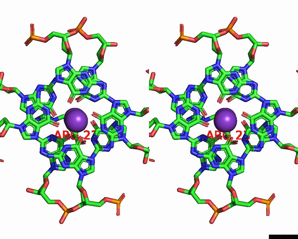
Stereo pair view
Mono view
Stereo pair view
A full contact list of Potassium with other atoms in the K binding
site number 1 of Crystal Structure of the Complex Between An Ankyrin and A Parallel G- Quadruplex within 5.0Å range:
|
Potassium binding site 2 out of 3 in 8ipp
Go back to
Potassium binding site 2 out
of 3 in the Crystal Structure of the Complex Between An Ankyrin and A Parallel G- Quadruplex
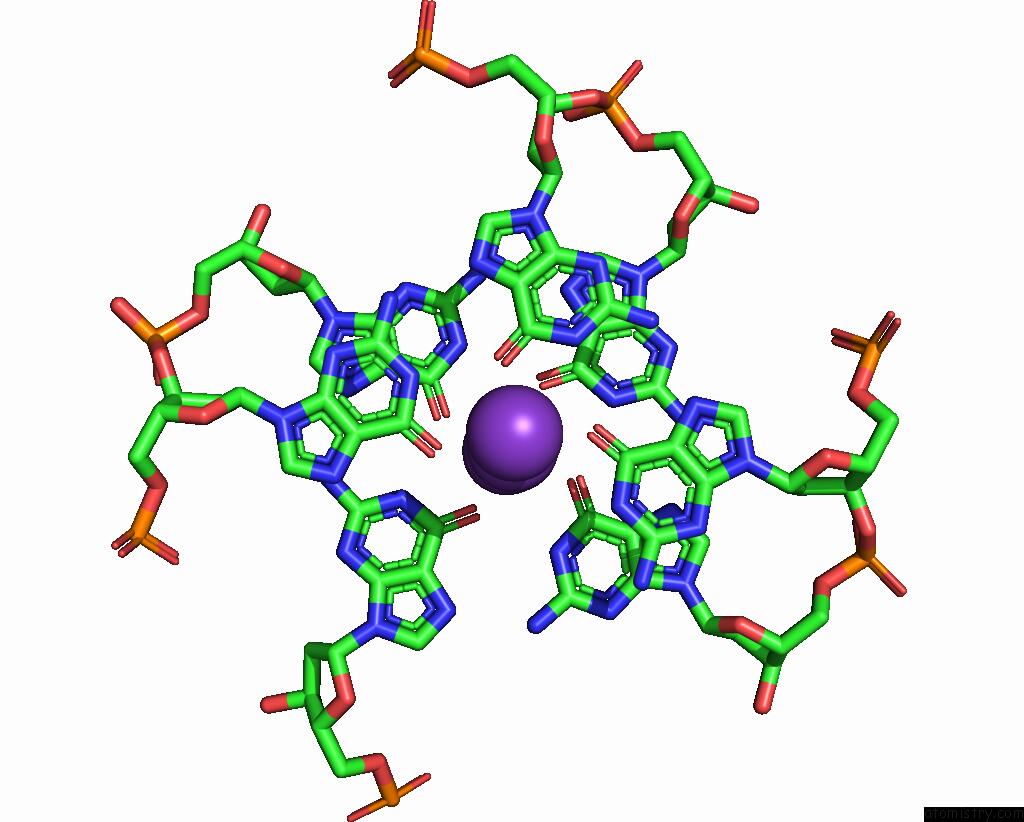
Mono view
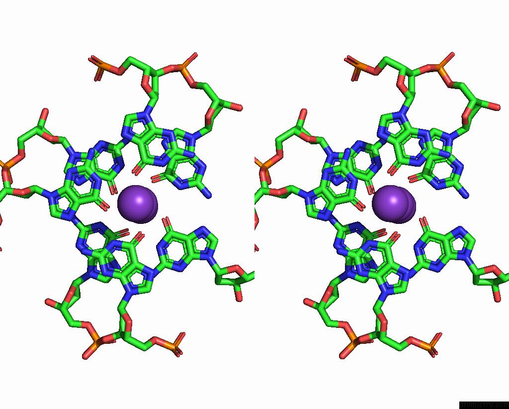
Stereo pair view
Mono view
Stereo pair view
A full contact list of Potassium with other atoms in the K binding
site number 2 of Crystal Structure of the Complex Between An Ankyrin and A Parallel G- Quadruplex within 5.0Å range:
|
Potassium binding site 3 out of 3 in 8ipp
Go back to
Potassium binding site 3 out
of 3 in the Crystal Structure of the Complex Between An Ankyrin and A Parallel G- Quadruplex
Mono view
Stereo pair view
Mono view
Stereo pair view
A full contact list of Potassium with other atoms in the K binding
site number 3 of Crystal Structure of the Complex Between An Ankyrin and A Parallel G- Quadruplex within 5.0Å range:
|
Reference:
K.H.Ngo,
C.W.Liew,
B.Heddi,
A.T.Phan.
Structural Basis For Parallel G-Quadruplex Recognition By An Ankyrin Protein. J.Am.Chem.Soc. V. 146 13709 2024.
ISSN: ESSN 1520-5126
PubMed: 38738955
DOI: 10.1021/JACS.4C01971
Page generated: Sat Aug 9 17:09:01 2025
ISSN: ESSN 1520-5126
PubMed: 38738955
DOI: 10.1021/JACS.4C01971
Last articles
Mg in 1L2OMg in 1L2E
Mg in 1L0O
Mg in 1L1R
Mg in 1KXG
Mg in 1KYR
Mg in 1KYP
Mg in 1KY3
Mg in 1KY2
Mg in 1KXP