Potassium »
PDB 8g7m-8jxo »
8gr9 »
Potassium in PDB 8gr9: Crystal Structure of Peroxisomal Citrate Synthase (CIT2) From Saccharomyces Cerevisiae in Complex with Oxaloacetate and Coenzyme-A
Protein crystallography data
The structure of Crystal Structure of Peroxisomal Citrate Synthase (CIT2) From Saccharomyces Cerevisiae in Complex with Oxaloacetate and Coenzyme-A, PDB code: 8gr9
was solved by
K.Nishio,
K.Nakatsukasa,
T.Kamura,
T.Mizushima,
with X-Ray Crystallography technique. A brief refinement statistics is given in the table below:
| Resolution Low / High (Å) | 34.13 / 1.48 |
| Space group | P 1 21 1 |
| Cell size a, b, c (Å), α, β, γ (°) | 63.079, 129.659, 68.127, 90, 116.57, 90 |
| R / Rfree (%) | 17.8 / 19.8 |
Other elements in 8gr9:
The structure of Crystal Structure of Peroxisomal Citrate Synthase (CIT2) From Saccharomyces Cerevisiae in Complex with Oxaloacetate and Coenzyme-A also contains other interesting chemical elements:
| Chlorine | (Cl) | 5 atoms |
Potassium Binding Sites:
The binding sites of Potassium atom in the Crystal Structure of Peroxisomal Citrate Synthase (CIT2) From Saccharomyces Cerevisiae in Complex with Oxaloacetate and Coenzyme-A
(pdb code 8gr9). This binding sites where shown within
5.0 Angstroms radius around Potassium atom.
In total 4 binding sites of Potassium where determined in the Crystal Structure of Peroxisomal Citrate Synthase (CIT2) From Saccharomyces Cerevisiae in Complex with Oxaloacetate and Coenzyme-A, PDB code: 8gr9:
Jump to Potassium binding site number: 1; 2; 3; 4;
In total 4 binding sites of Potassium where determined in the Crystal Structure of Peroxisomal Citrate Synthase (CIT2) From Saccharomyces Cerevisiae in Complex with Oxaloacetate and Coenzyme-A, PDB code: 8gr9:
Jump to Potassium binding site number: 1; 2; 3; 4;
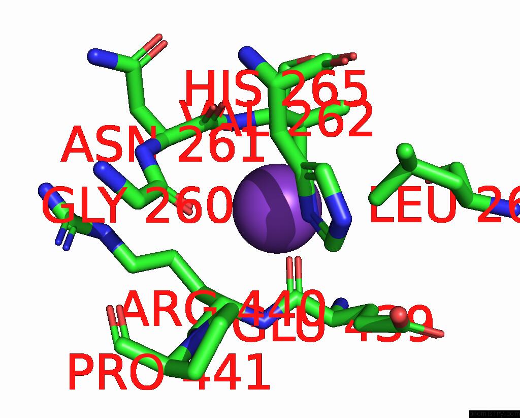
Potassium binding site 1 out of 4 in 8gr9
Go back to
Potassium binding site 1 out
of 4 in the Crystal Structure of Peroxisomal Citrate Synthase (CIT2) From Saccharomyces Cerevisiae in Complex with Oxaloacetate and Coenzyme-A
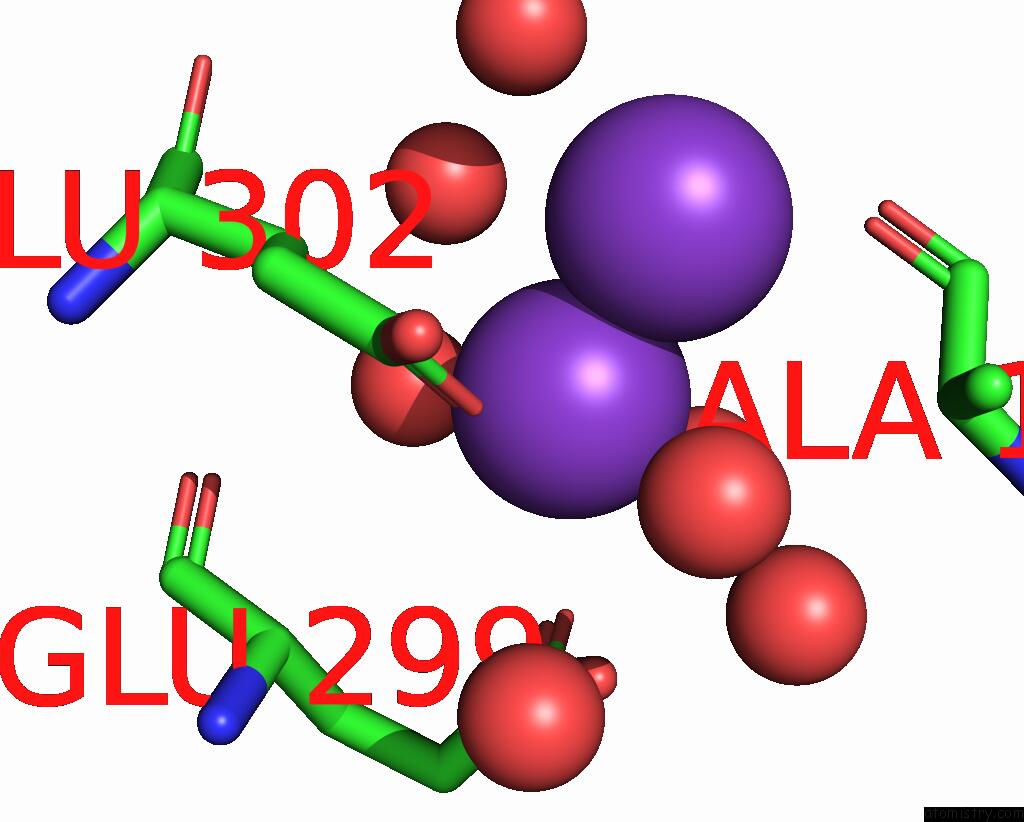
Mono view
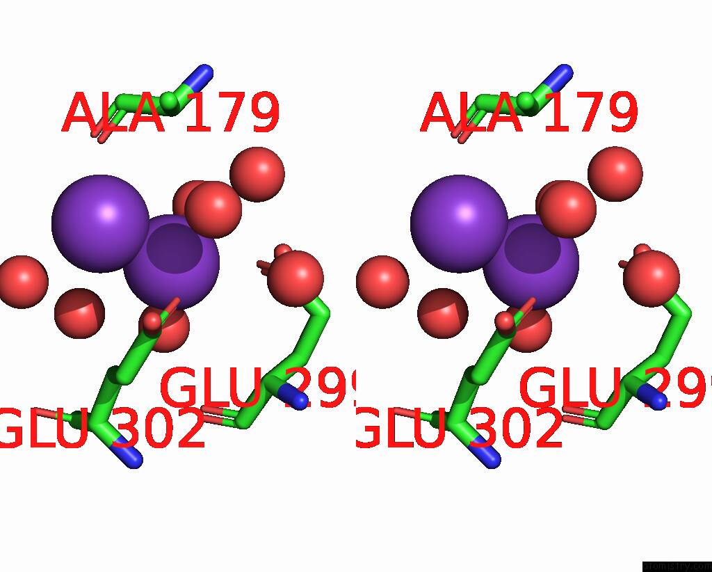
Stereo pair view
Mono view
Stereo pair view
A full contact list of Potassium with other atoms in the K binding
site number 1 of Crystal Structure of Peroxisomal Citrate Synthase (CIT2) From Saccharomyces Cerevisiae in Complex with Oxaloacetate and Coenzyme-A within 5.0Å range:
|
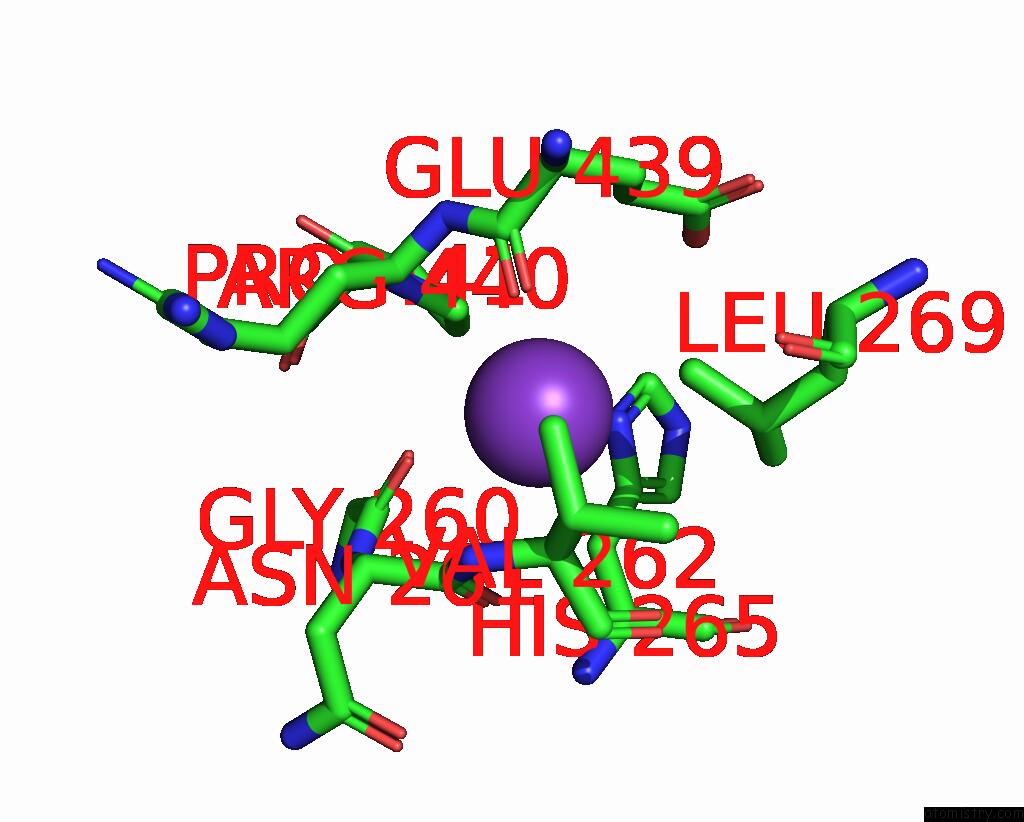
Potassium binding site 2 out of 4 in 8gr9
Go back to
Potassium binding site 2 out
of 4 in the Crystal Structure of Peroxisomal Citrate Synthase (CIT2) From Saccharomyces Cerevisiae in Complex with Oxaloacetate and Coenzyme-A
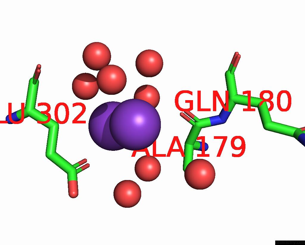
Mono view
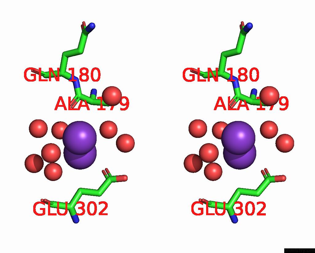
Stereo pair view
Mono view
Stereo pair view
A full contact list of Potassium with other atoms in the K binding
site number 2 of Crystal Structure of Peroxisomal Citrate Synthase (CIT2) From Saccharomyces Cerevisiae in Complex with Oxaloacetate and Coenzyme-A within 5.0Å range:
|
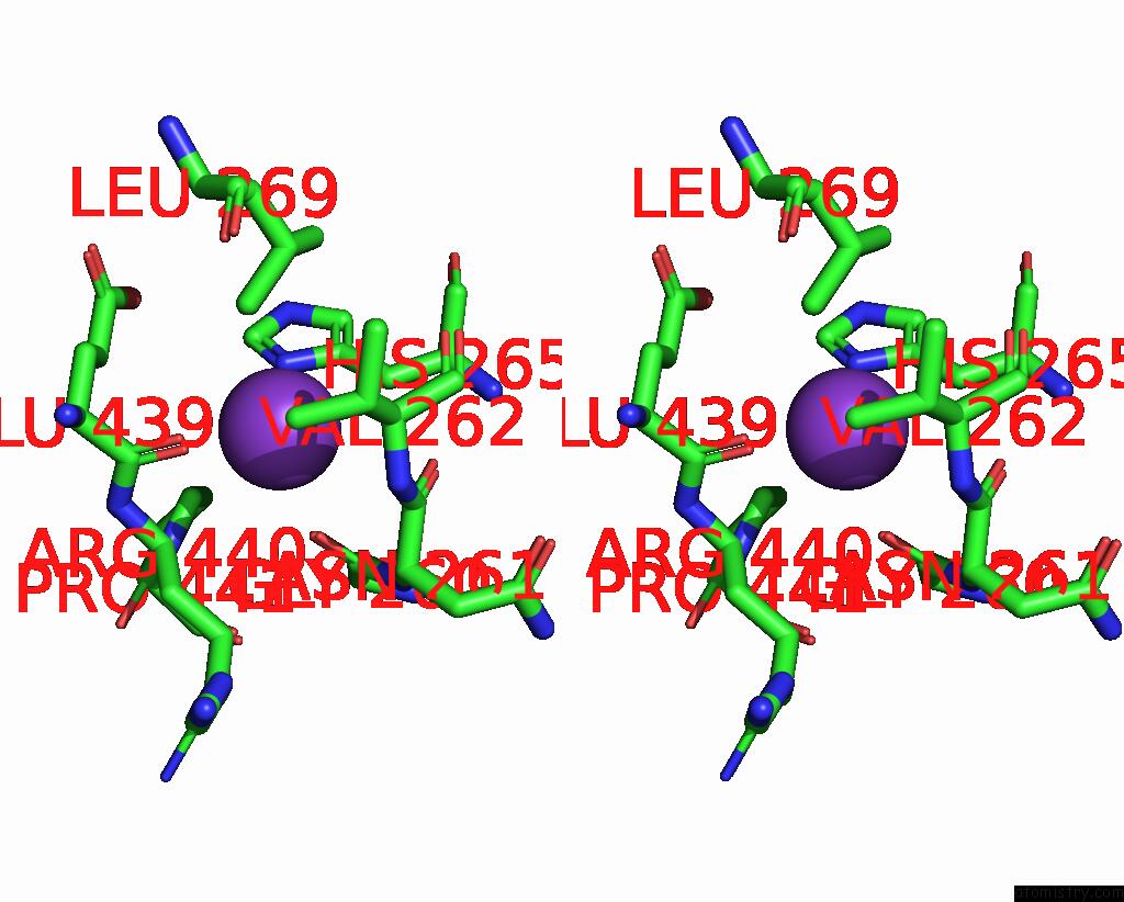
Potassium binding site 3 out of 4 in 8gr9
Go back to
Potassium binding site 3 out
of 4 in the Crystal Structure of Peroxisomal Citrate Synthase (CIT2) From Saccharomyces Cerevisiae in Complex with Oxaloacetate and Coenzyme-A
Mono view

Stereo pair view
Mono view
Stereo pair view
A full contact list of Potassium with other atoms in the K binding
site number 3 of Crystal Structure of Peroxisomal Citrate Synthase (CIT2) From Saccharomyces Cerevisiae in Complex with Oxaloacetate and Coenzyme-A within 5.0Å range:
|
Potassium binding site 4 out of 4 in 8gr9
Go back to
Potassium binding site 4 out
of 4 in the Crystal Structure of Peroxisomal Citrate Synthase (CIT2) From Saccharomyces Cerevisiae in Complex with Oxaloacetate and Coenzyme-A
Mono view
Stereo pair view
Mono view
Stereo pair view
A full contact list of Potassium with other atoms in the K binding
site number 4 of Crystal Structure of Peroxisomal Citrate Synthase (CIT2) From Saccharomyces Cerevisiae in Complex with Oxaloacetate and Coenzyme-A within 5.0Å range:
|
Reference:
K.Nishio,
T.Kawarasaki,
Y.Sugiura,
S.Matsumoto,
A.Konoshima,
Y.Takano,
M.Hayashi,
F.Okumura,
T.Kamura,
T.Mizushima,
K.Nakatsukasa.
Defective Import of Mitochondrial Metabolic Enzyme Elicits Ectopic Metabolic Stress. Sci Adv V. 9 F1956 2023.
ISSN: ESSN 2375-2548
PubMed: 37058555
DOI: 10.1126/SCIADV.ADF1956
Page generated: Mon Aug 12 23:52:17 2024
ISSN: ESSN 2375-2548
PubMed: 37058555
DOI: 10.1126/SCIADV.ADF1956
Last articles
Zn in 9JYWZn in 9IR4
Zn in 9IR3
Zn in 9GMX
Zn in 9GMW
Zn in 9JEJ
Zn in 9ERF
Zn in 9ERE
Zn in 9EGV
Zn in 9EGW