Potassium »
PDB 8g7m-8jxo »
8gpe »
Potassium in PDB 8gpe: Crystal Structure of Ndm-1 at PH5.5 (Succinate) in Complex with Hydrolyzed Penicillin G
Protein crystallography data
The structure of Crystal Structure of Ndm-1 at PH5.5 (Succinate) in Complex with Hydrolyzed Penicillin G, PDB code: 8gpe
was solved by
X.Shi,
Y.Dai,
Q.Zhang,
W.Liu,
with X-Ray Crystallography technique. A brief refinement statistics is given in the table below:
| Resolution Low / High (Å) | 38.93 / 1.40 |
| Space group | P 21 21 21 |
| Cell size a, b, c (Å), α, β, γ (°) | 39.17, 79.16, 134.12, 90, 90, 90 |
| R / Rfree (%) | 13 / 16 |
Other elements in 8gpe:
The structure of Crystal Structure of Ndm-1 at PH5.5 (Succinate) in Complex with Hydrolyzed Penicillin G also contains other interesting chemical elements:
| Zinc | (Zn) | 4 atoms |
Potassium Binding Sites:
The binding sites of Potassium atom in the Crystal Structure of Ndm-1 at PH5.5 (Succinate) in Complex with Hydrolyzed Penicillin G
(pdb code 8gpe). This binding sites where shown within
5.0 Angstroms radius around Potassium atom.
In total only one binding site of Potassium was determined in the Crystal Structure of Ndm-1 at PH5.5 (Succinate) in Complex with Hydrolyzed Penicillin G, PDB code: 8gpe:
In total only one binding site of Potassium was determined in the Crystal Structure of Ndm-1 at PH5.5 (Succinate) in Complex with Hydrolyzed Penicillin G, PDB code: 8gpe:
Potassium binding site 1 out of 1 in 8gpe
Go back to
Potassium binding site 1 out
of 1 in the Crystal Structure of Ndm-1 at PH5.5 (Succinate) in Complex with Hydrolyzed Penicillin G
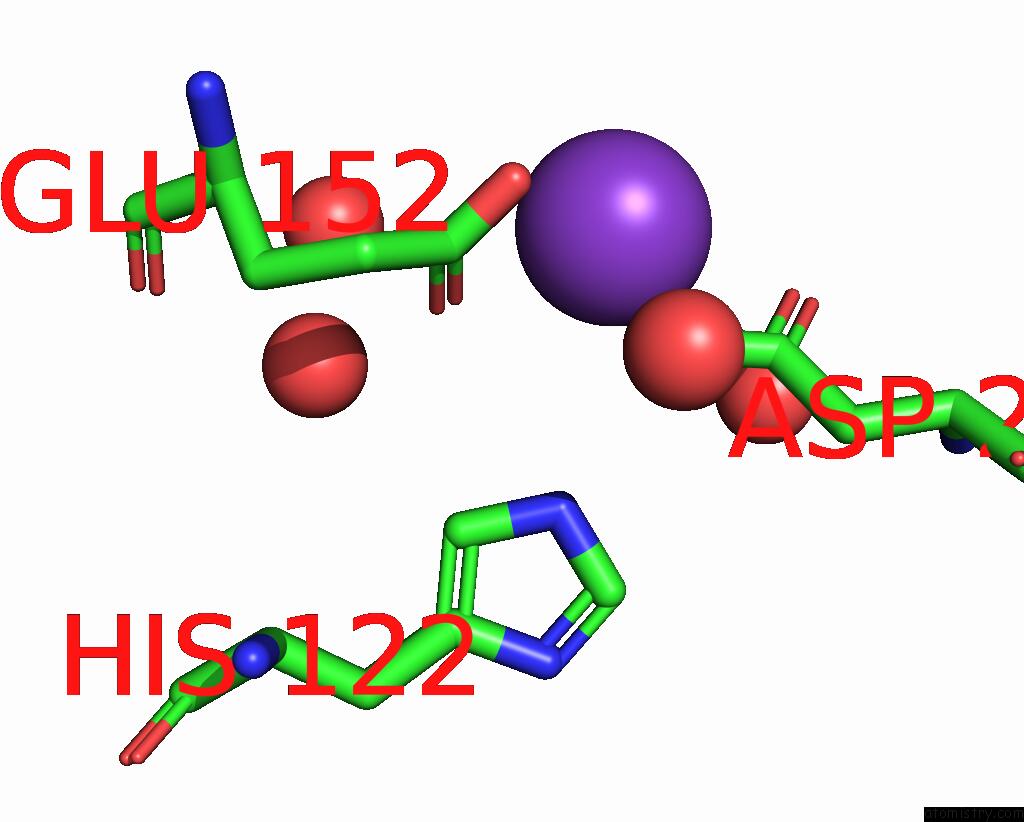
Mono view
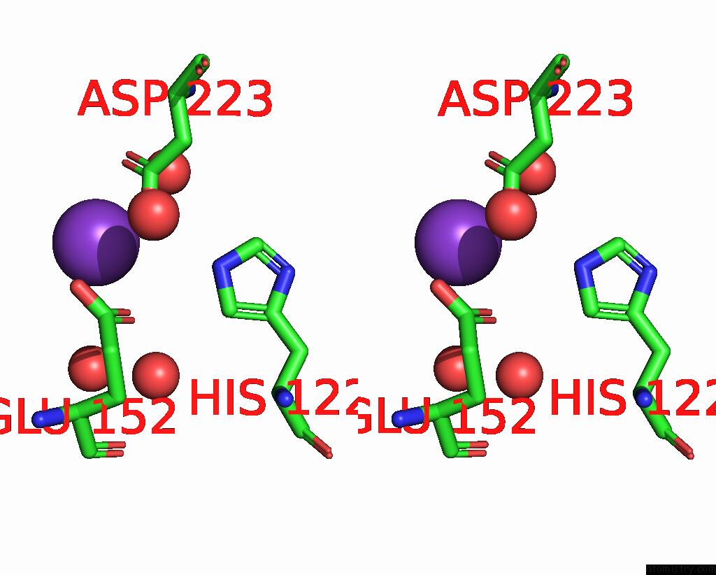
Stereo pair view
Mono view
Stereo pair view
A full contact list of Potassium with other atoms in the K binding
site number 1 of Crystal Structure of Ndm-1 at PH5.5 (Succinate) in Complex with Hydrolyzed Penicillin G within 5.0Å range:
|
Reference:
X.Shi,
Y.Dai,
Q.Zhang,
W.Liu.
Crystal Structure of Ndm-1 at PH5.5 (Succinate) in Complex with Hydrolyzed Penicillin G To Be Published.
Page generated: Mon Aug 12 23:52:02 2024
Last articles
Zn in 9JYWZn in 9IR4
Zn in 9IR3
Zn in 9GMX
Zn in 9GMW
Zn in 9JEJ
Zn in 9ERF
Zn in 9ERE
Zn in 9EGV
Zn in 9EGW