Potassium in PDB 8evx: Ddla From Pseudomonas Aeruginosa PAO1 in Complex with Adp and Phosphorylated D-Cycloserine
Enzymatic activity of Ddla From Pseudomonas Aeruginosa PAO1 in Complex with Adp and Phosphorylated D-Cycloserine
All present enzymatic activity of Ddla From Pseudomonas Aeruginosa PAO1 in Complex with Adp and Phosphorylated D-Cycloserine:
6.3.2.4;
6.3.2.4;
Protein crystallography data
The structure of Ddla From Pseudomonas Aeruginosa PAO1 in Complex with Adp and Phosphorylated D-Cycloserine, PDB code: 8evx
was solved by
J.L.Pederick,
J.C.Woolman,
J.B.Bruning,
with X-Ray Crystallography technique. A brief refinement statistics is given in the table below:
| Resolution Low / High (Å) | 37.18 / 1.55 |
| Space group | P 1 21 1 |
| Cell size a, b, c (Å), α, β, γ (°) | 47.467, 133.053, 53.852, 90, 109.17, 90 |
| R / Rfree (%) | 19.8 / 21.8 |
Other elements in 8evx:
The structure of Ddla From Pseudomonas Aeruginosa PAO1 in Complex with Adp and Phosphorylated D-Cycloserine also contains other interesting chemical elements:
| Magnesium | (Mg) | 4 atoms |
Potassium Binding Sites:
The binding sites of Potassium atom in the Ddla From Pseudomonas Aeruginosa PAO1 in Complex with Adp and Phosphorylated D-Cycloserine
(pdb code 8evx). This binding sites where shown within
5.0 Angstroms radius around Potassium atom.
In total 2 binding sites of Potassium where determined in the Ddla From Pseudomonas Aeruginosa PAO1 in Complex with Adp and Phosphorylated D-Cycloserine, PDB code: 8evx:
Jump to Potassium binding site number: 1; 2;
In total 2 binding sites of Potassium where determined in the Ddla From Pseudomonas Aeruginosa PAO1 in Complex with Adp and Phosphorylated D-Cycloserine, PDB code: 8evx:
Jump to Potassium binding site number: 1; 2;
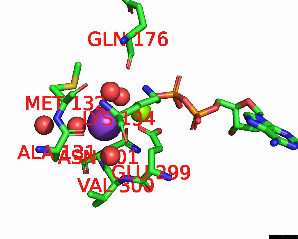
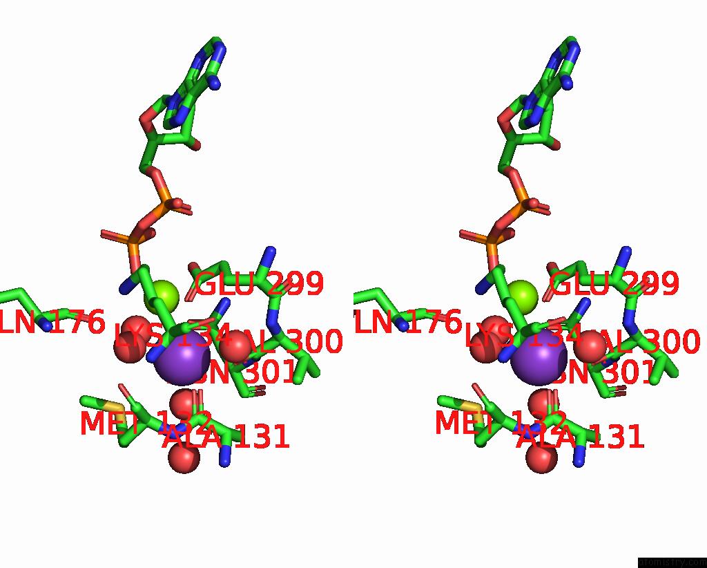
Potassium binding site 1 out of 2 in 8evx
Go back to
Potassium binding site 1 out
of 2 in the Ddla From Pseudomonas Aeruginosa PAO1 in Complex with Adp and Phosphorylated D-Cycloserine

Mono view

Stereo pair view
Mono view
Stereo pair view
A full contact list of Potassium with other atoms in the K binding
site number 1 of Ddla From Pseudomonas Aeruginosa PAO1 in Complex with Adp and Phosphorylated D-Cycloserine within 5.0Å range:
|
Potassium binding site 2 out of 2 in 8evx
Go back to
Potassium binding site 2 out
of 2 in the Ddla From Pseudomonas Aeruginosa PAO1 in Complex with Adp and Phosphorylated D-Cycloserine
Mono view
Stereo pair view
Mono view
Stereo pair view
A full contact list of Potassium with other atoms in the K binding
site number 2 of Ddla From Pseudomonas Aeruginosa PAO1 in Complex with Adp and Phosphorylated D-Cycloserine within 5.0Å range:
|
Reference:
J.L.Pederick,
J.C.Woolman,
J.B.Bruning.
Comparative Functional and Structural Analysis of Pseudomonas Aeruginosa ᴅ-Alanine-ᴅ-Alanine Ligase Isoforms As Prospective Antibiotic Targets. Febs J. 2023.
ISSN: ISSN 1742-464X
PubMed: 37581574
DOI: 10.1111/FEBS.16932
Page generated: Mon Aug 12 23:36:42 2024
ISSN: ISSN 1742-464X
PubMed: 37581574
DOI: 10.1111/FEBS.16932
Last articles
Zn in 9MJ5Zn in 9HNW
Zn in 9G0L
Zn in 9FNE
Zn in 9DZN
Zn in 9E0I
Zn in 9D32
Zn in 9DAK
Zn in 8ZXC
Zn in 8ZUF