Potassium »
PDB 8ctu-8djb »
8df9 »
Potassium in PDB 8df9: Structure of M. Kandleri Topoisomerase V in Complex with Dna. 38 Base Pair Asymmetric Dna Complex
Protein crystallography data
The structure of Structure of M. Kandleri Topoisomerase V in Complex with Dna. 38 Base Pair Asymmetric Dna Complex, PDB code: 8df9
was solved by
A.Osterman,
A.Mondragon,
with X-Ray Crystallography technique. A brief refinement statistics is given in the table below:
| Resolution Low / High (Å) | 39.28 / 3.24 |
| Space group | P 41 21 2 |
| Cell size a, b, c (Å), α, β, γ (°) | 193.751, 193.751, 245.979, 90, 90, 90 |
| R / Rfree (%) | 23.5 / 26.6 |
Other elements in 8df9:
The structure of Structure of M. Kandleri Topoisomerase V in Complex with Dna. 38 Base Pair Asymmetric Dna Complex also contains other interesting chemical elements:
| Magnesium | (Mg) | 12 atoms |
Potassium Binding Sites:
The binding sites of Potassium atom in the Structure of M. Kandleri Topoisomerase V in Complex with Dna. 38 Base Pair Asymmetric Dna Complex
(pdb code 8df9). This binding sites where shown within
5.0 Angstroms radius around Potassium atom.
In total 2 binding sites of Potassium where determined in the Structure of M. Kandleri Topoisomerase V in Complex with Dna. 38 Base Pair Asymmetric Dna Complex, PDB code: 8df9:
Jump to Potassium binding site number: 1; 2;
In total 2 binding sites of Potassium where determined in the Structure of M. Kandleri Topoisomerase V in Complex with Dna. 38 Base Pair Asymmetric Dna Complex, PDB code: 8df9:
Jump to Potassium binding site number: 1; 2;
Potassium binding site 1 out of 2 in 8df9
Go back to
Potassium binding site 1 out
of 2 in the Structure of M. Kandleri Topoisomerase V in Complex with Dna. 38 Base Pair Asymmetric Dna Complex
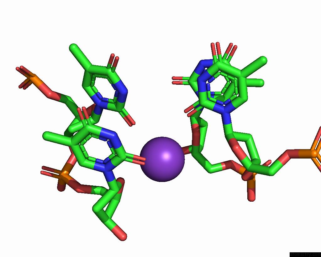
Mono view
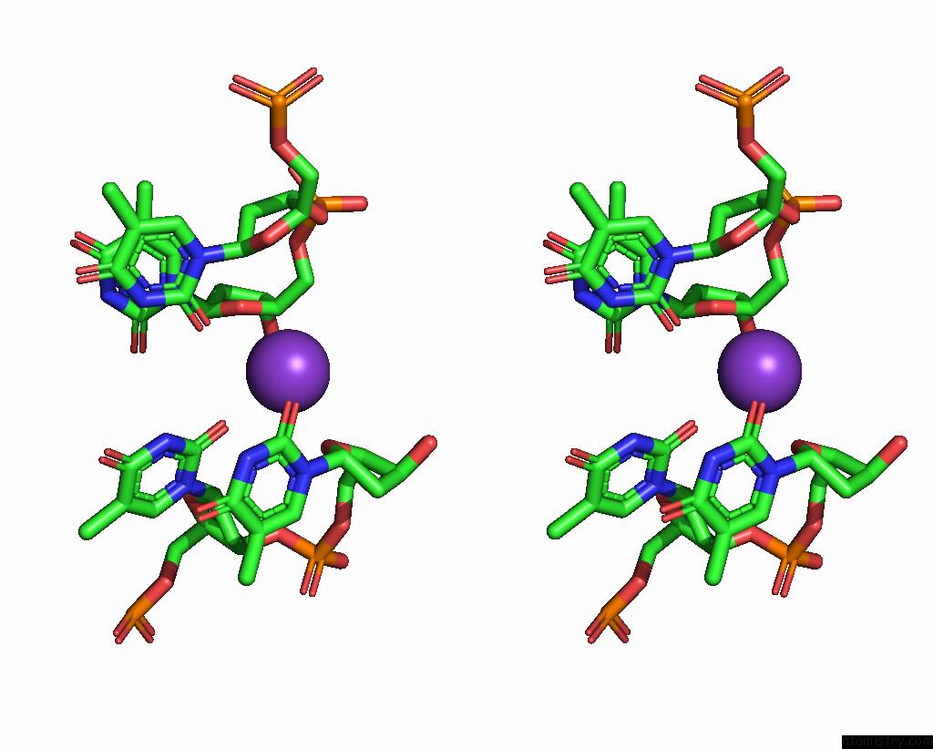
Stereo pair view
Mono view
Stereo pair view
A full contact list of Potassium with other atoms in the K binding
site number 1 of Structure of M. Kandleri Topoisomerase V in Complex with Dna. 38 Base Pair Asymmetric Dna Complex within 5.0Å range:
|
Potassium binding site 2 out of 2 in 8df9
Go back to
Potassium binding site 2 out
of 2 in the Structure of M. Kandleri Topoisomerase V in Complex with Dna. 38 Base Pair Asymmetric Dna Complex
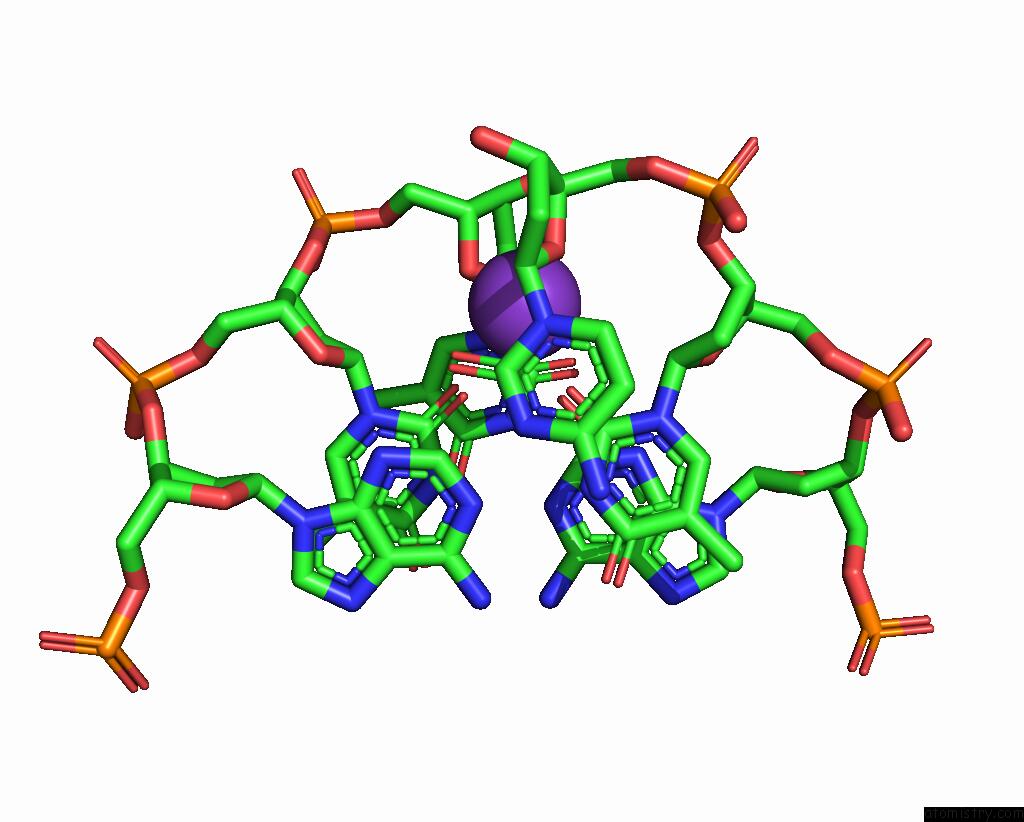
Mono view
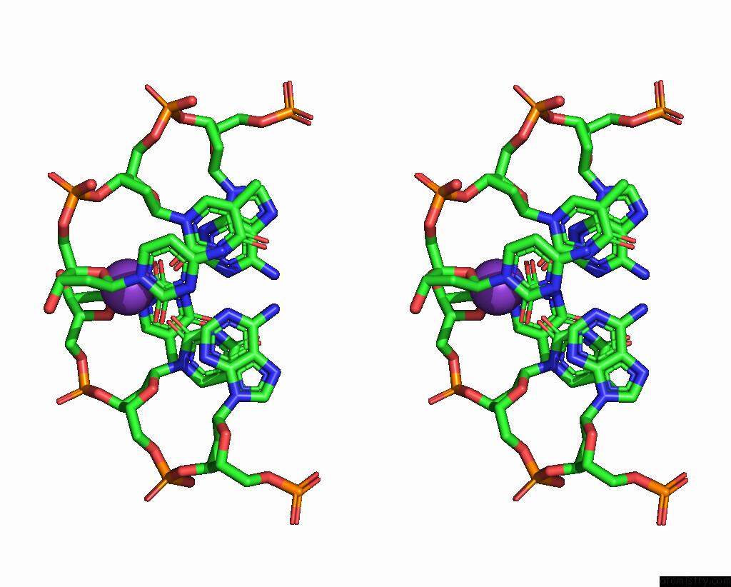
Stereo pair view
Mono view
Stereo pair view
A full contact list of Potassium with other atoms in the K binding
site number 2 of Structure of M. Kandleri Topoisomerase V in Complex with Dna. 38 Base Pair Asymmetric Dna Complex within 5.0Å range:
|
Reference:
A.Osterman,
A.Mondragon.
Structures of Topoisomerase V in Complex with Dna Reveal Unusual Dna Binding Mode and Novel Relaxation Mechanism. Elife V. 11 2022.
ISSN: ESSN 2050-084X
PubMed: 35969036
DOI: 10.7554/ELIFE.72702
Page generated: Sat Aug 9 16:43:54 2025
ISSN: ESSN 2050-084X
PubMed: 35969036
DOI: 10.7554/ELIFE.72702
Last articles
Mg in 5JWAMg in 5JVL
Mg in 5JVN
Mg in 5JVM
Mg in 5JVJ
Mg in 5JVD
Mg in 5JV5
Mg in 5JTG
Mg in 5JRW
Mg in 5JSQ