Potassium »
PDB 8cfh-8ctt »
8cfy »
Potassium in PDB 8cfy: Crystal Structure of S-Adenosyl-L-Homocysteine Hydrolase From P. Aeruginosa in Complex with F2X-Entry Library Fragment H08
Enzymatic activity of Crystal Structure of S-Adenosyl-L-Homocysteine Hydrolase From P. Aeruginosa in Complex with F2X-Entry Library Fragment H08
All present enzymatic activity of Crystal Structure of S-Adenosyl-L-Homocysteine Hydrolase From P. Aeruginosa in Complex with F2X-Entry Library Fragment H08:
3.3.1.1;
3.3.1.1;
Protein crystallography data
The structure of Crystal Structure of S-Adenosyl-L-Homocysteine Hydrolase From P. Aeruginosa in Complex with F2X-Entry Library Fragment H08, PDB code: 8cfy
was solved by
P.H.Malecki,
M.Gawel,
M.Stepniewska,
K.Brzezinski,
with X-Ray Crystallography technique. A brief refinement statistics is given in the table below:
| Resolution Low / High (Å) | 42.61 / 1.88 |
| Space group | P 1 2 1 |
| Cell size a, b, c (Å), α, β, γ (°) | 109.085, 103.191, 175.392, 90, 99.55, 90 |
| R / Rfree (%) | 17.4 / 20.9 |
Potassium Binding Sites:
The binding sites of Potassium atom in the Crystal Structure of S-Adenosyl-L-Homocysteine Hydrolase From P. Aeruginosa in Complex with F2X-Entry Library Fragment H08
(pdb code 8cfy). This binding sites where shown within
5.0 Angstroms radius around Potassium atom.
In total 8 binding sites of Potassium where determined in the Crystal Structure of S-Adenosyl-L-Homocysteine Hydrolase From P. Aeruginosa in Complex with F2X-Entry Library Fragment H08, PDB code: 8cfy:
Jump to Potassium binding site number: 1; 2; 3; 4; 5; 6; 7; 8;
In total 8 binding sites of Potassium where determined in the Crystal Structure of S-Adenosyl-L-Homocysteine Hydrolase From P. Aeruginosa in Complex with F2X-Entry Library Fragment H08, PDB code: 8cfy:
Jump to Potassium binding site number: 1; 2; 3; 4; 5; 6; 7; 8;
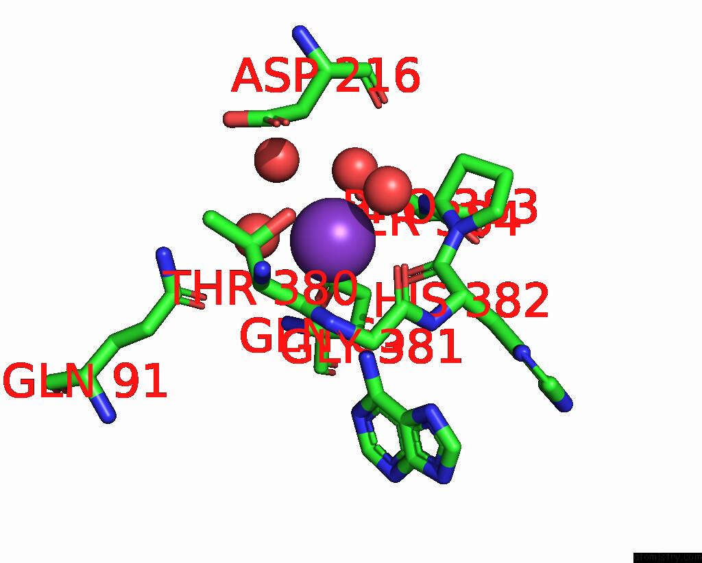
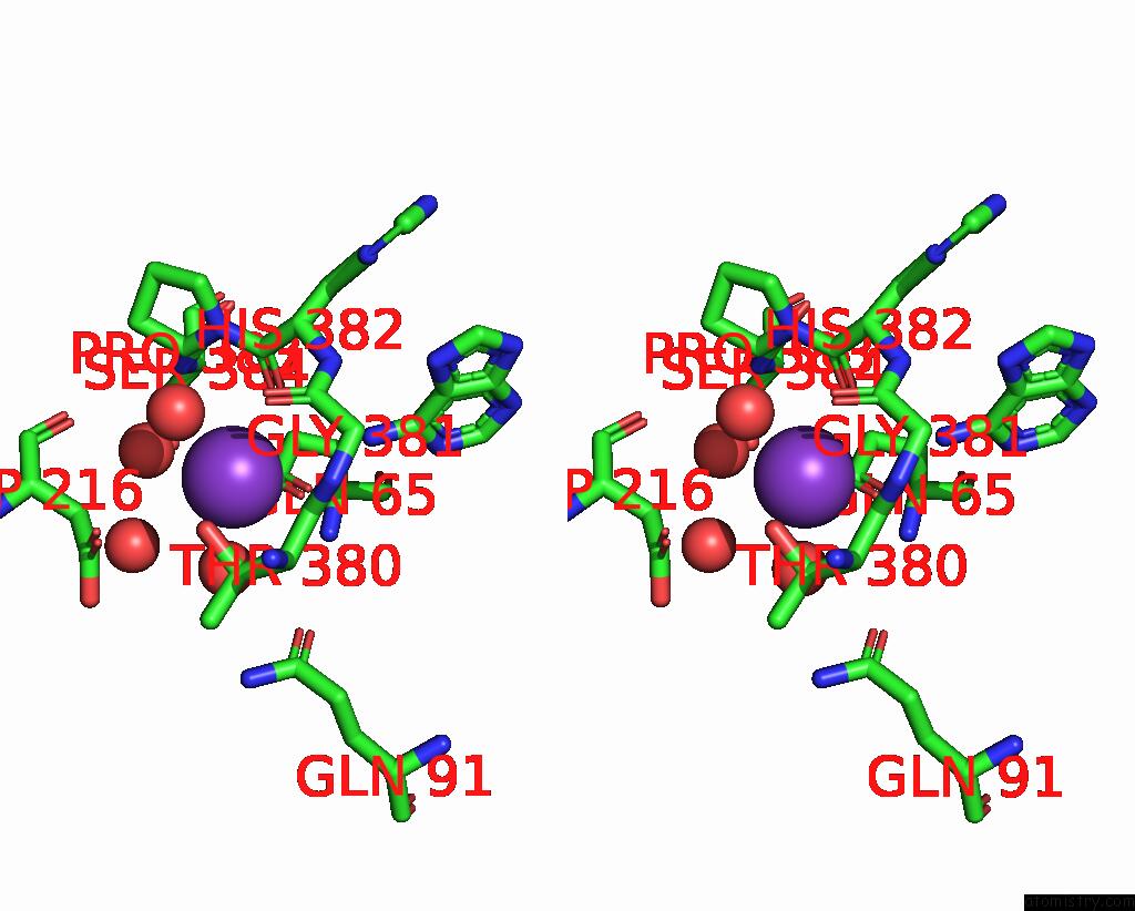
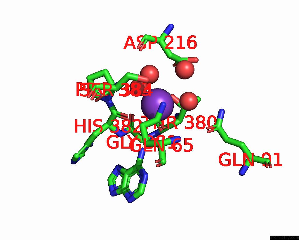
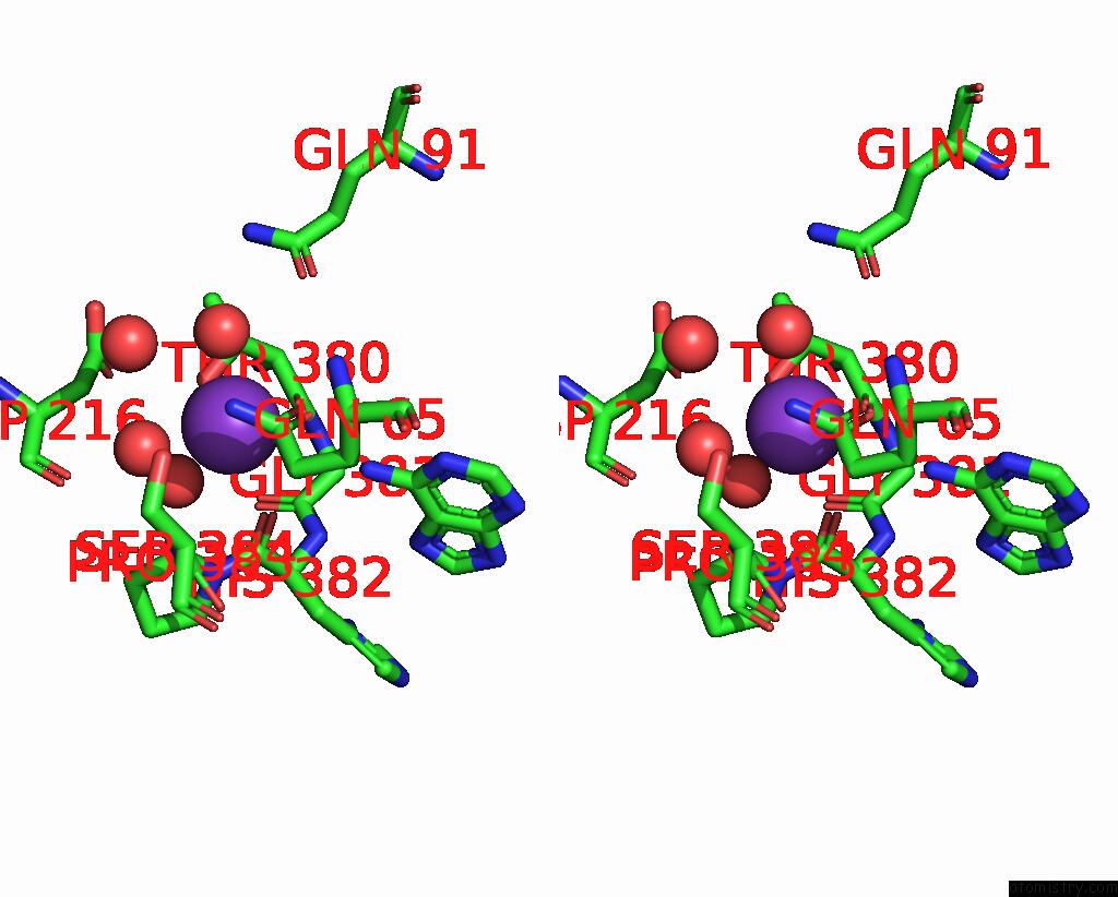
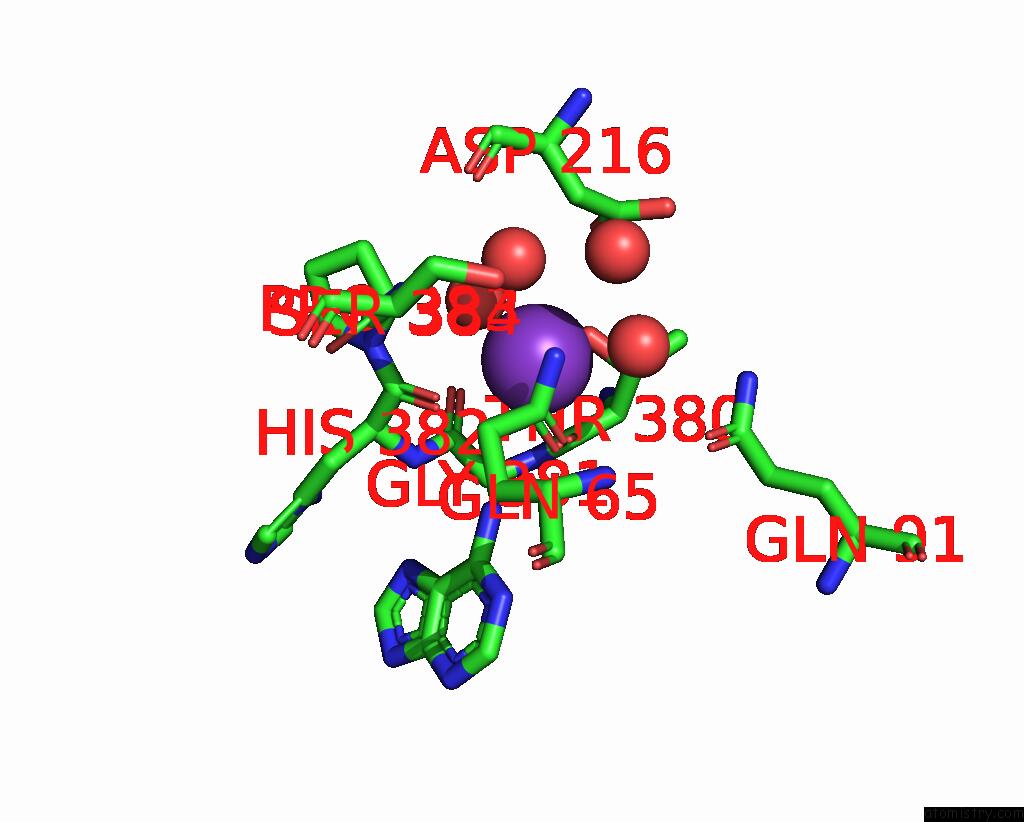
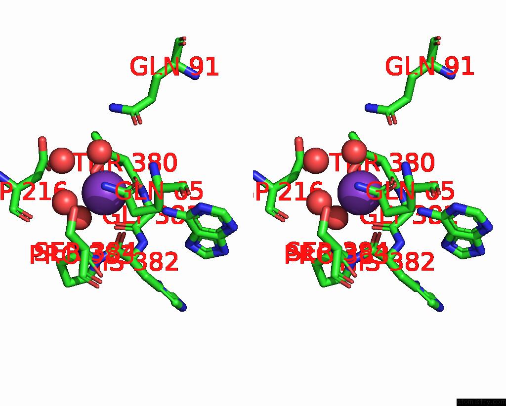
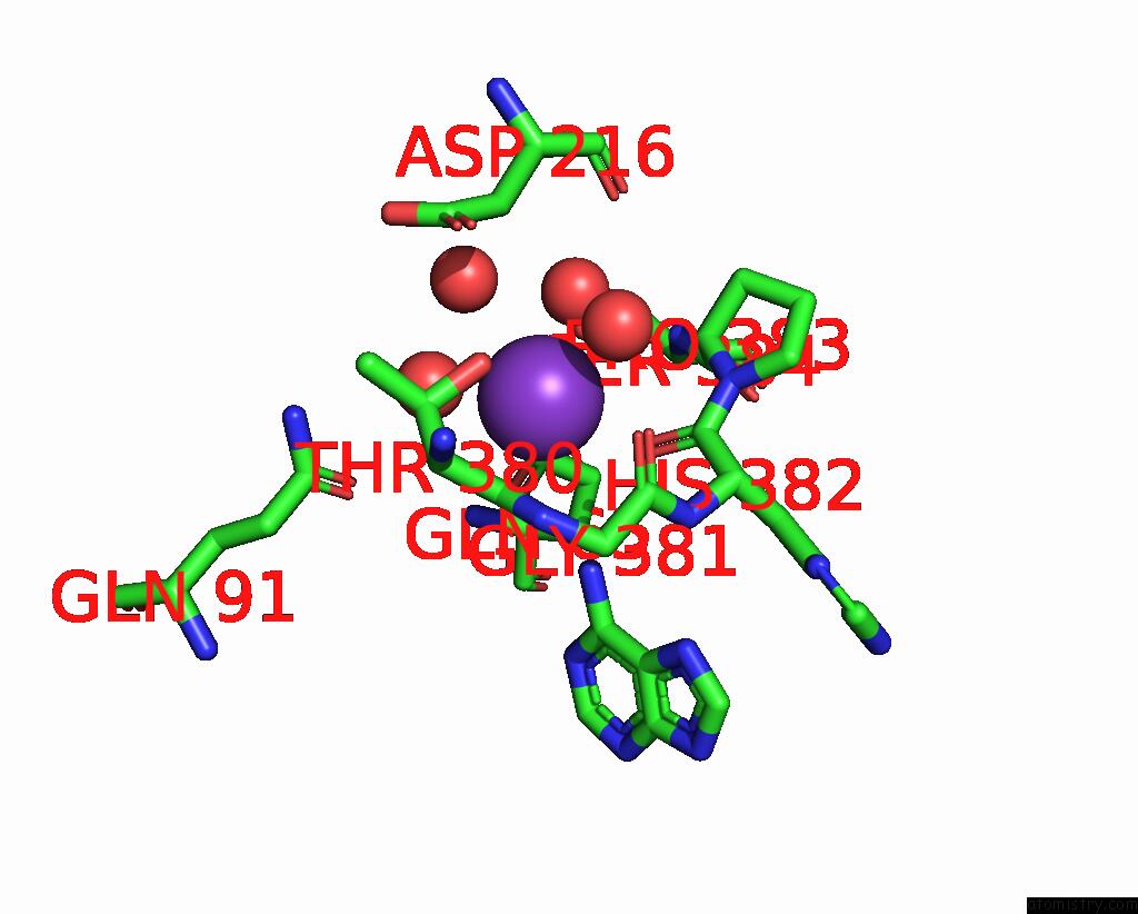
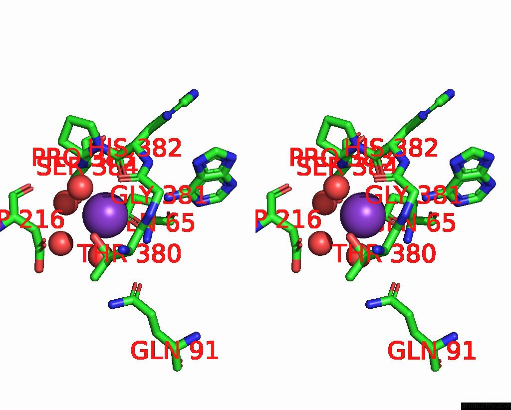
Potassium binding site 1 out of 8 in 8cfy
Go back to
Potassium binding site 1 out
of 8 in the Crystal Structure of S-Adenosyl-L-Homocysteine Hydrolase From P. Aeruginosa in Complex with F2X-Entry Library Fragment H08
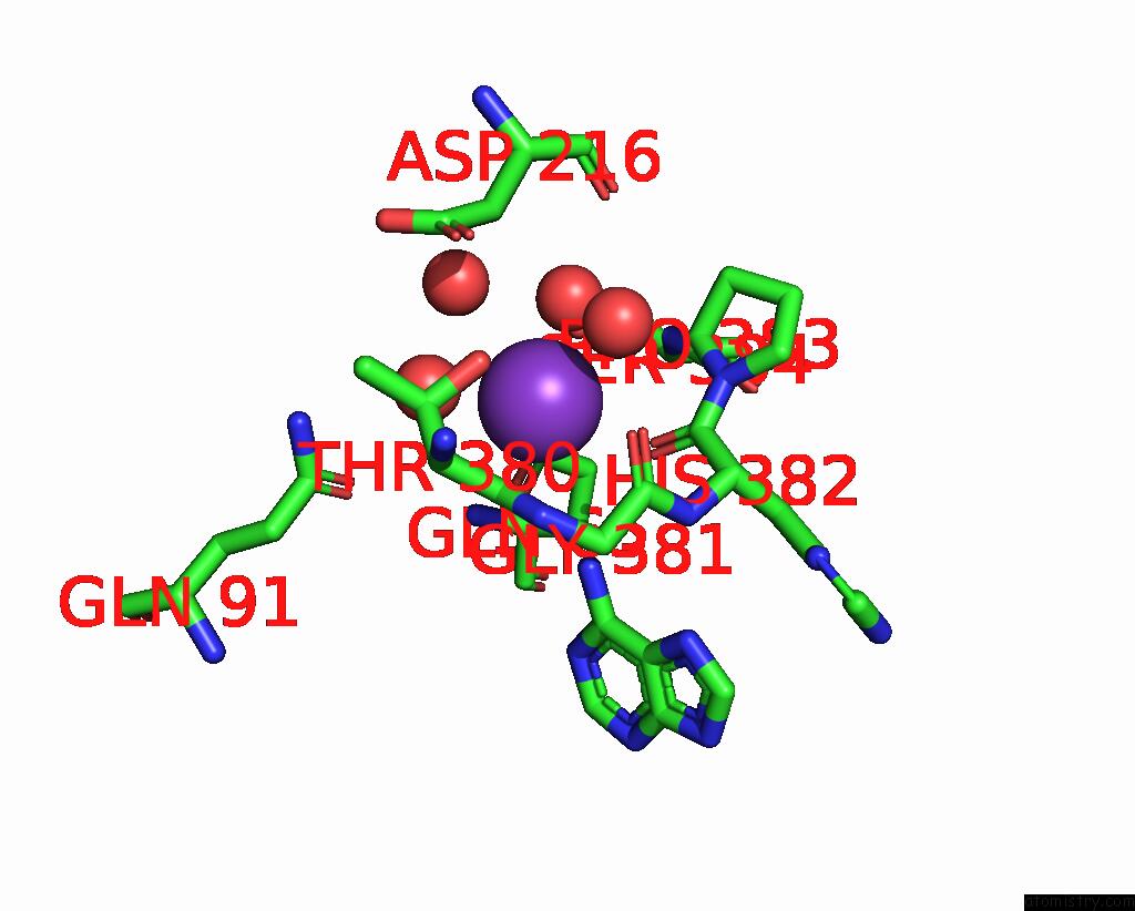
Mono view
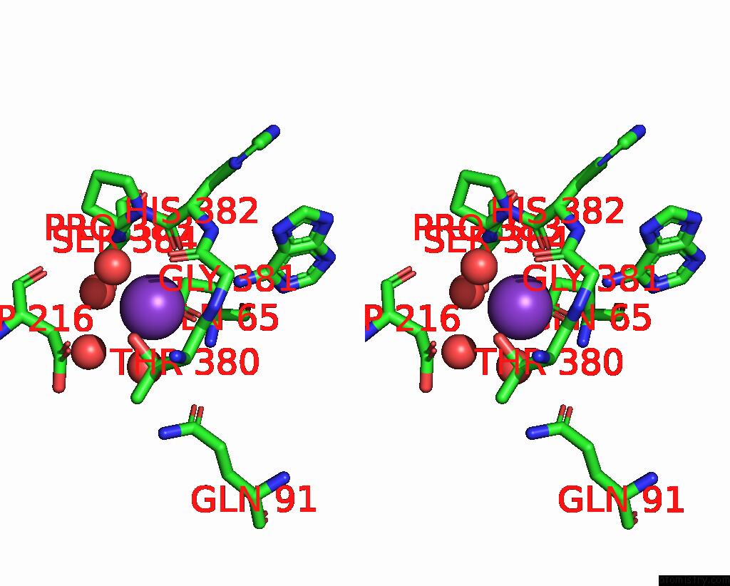
Stereo pair view
Mono view
Stereo pair view
A full contact list of Potassium with other atoms in the K binding
site number 1 of Crystal Structure of S-Adenosyl-L-Homocysteine Hydrolase From P. Aeruginosa in Complex with F2X-Entry Library Fragment H08 within 5.0Å range:
|
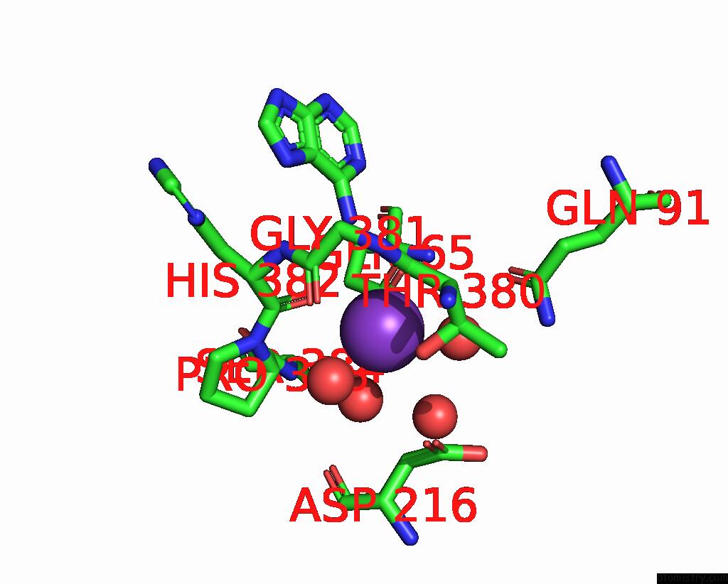
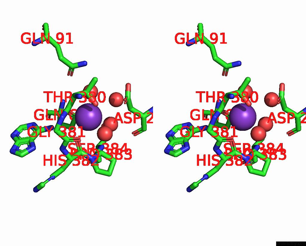
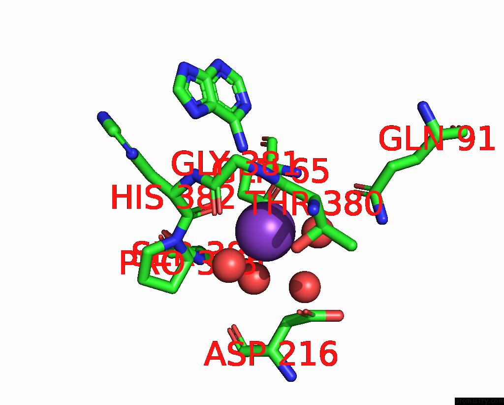
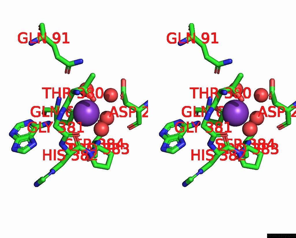
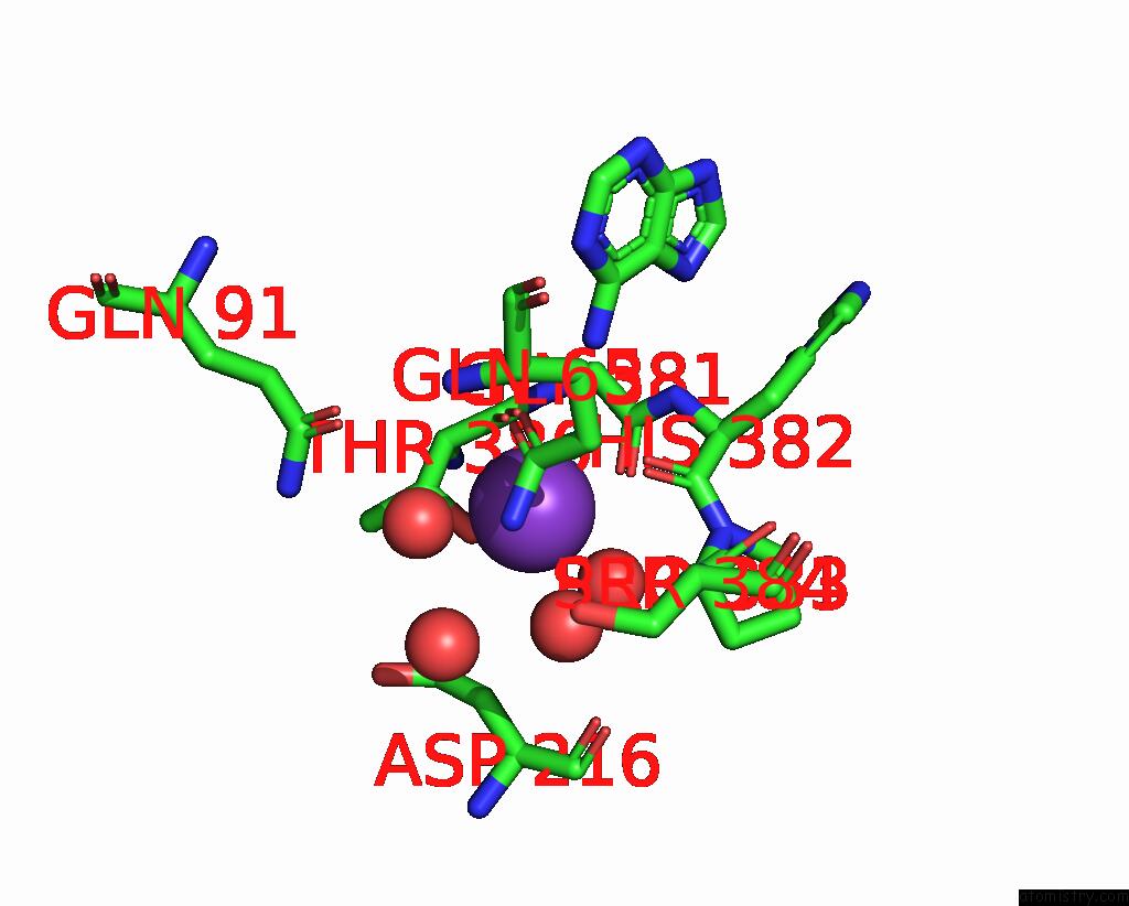
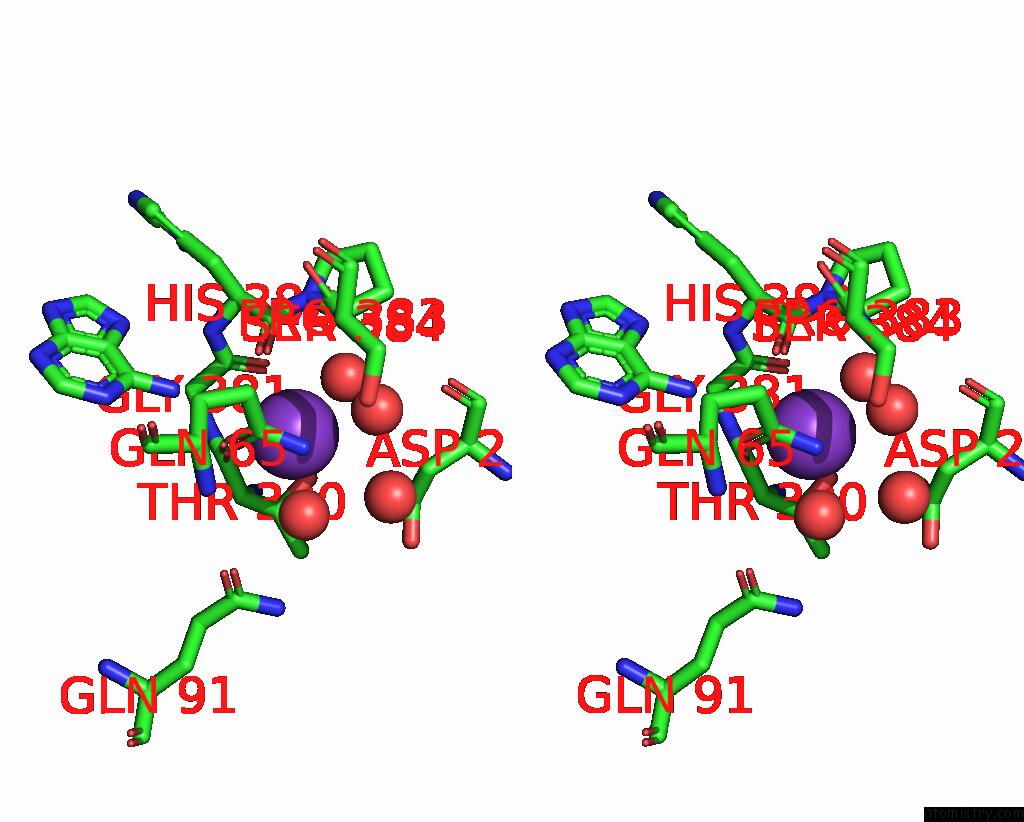
Potassium binding site 2 out of 8 in 8cfy
Go back to
Potassium binding site 2 out
of 8 in the Crystal Structure of S-Adenosyl-L-Homocysteine Hydrolase From P. Aeruginosa in Complex with F2X-Entry Library Fragment H08
Mono view
Stereo pair view
Mono view
Stereo pair view
A full contact list of Potassium with other atoms in the K binding
site number 2 of Crystal Structure of S-Adenosyl-L-Homocysteine Hydrolase From P. Aeruginosa in Complex with F2X-Entry Library Fragment H08 within 5.0Å range:
|
Potassium binding site 3 out of 8 in 8cfy
Go back to
Potassium binding site 3 out
of 8 in the Crystal Structure of S-Adenosyl-L-Homocysteine Hydrolase From P. Aeruginosa in Complex with F2X-Entry Library Fragment H08
Mono view
Stereo pair view
Mono view
Stereo pair view
A full contact list of Potassium with other atoms in the K binding
site number 3 of Crystal Structure of S-Adenosyl-L-Homocysteine Hydrolase From P. Aeruginosa in Complex with F2X-Entry Library Fragment H08 within 5.0Å range:
|
Potassium binding site 4 out of 8 in 8cfy
Go back to
Potassium binding site 4 out
of 8 in the Crystal Structure of S-Adenosyl-L-Homocysteine Hydrolase From P. Aeruginosa in Complex with F2X-Entry Library Fragment H08
Mono view
Stereo pair view
Mono view
Stereo pair view
A full contact list of Potassium with other atoms in the K binding
site number 4 of Crystal Structure of S-Adenosyl-L-Homocysteine Hydrolase From P. Aeruginosa in Complex with F2X-Entry Library Fragment H08 within 5.0Å range:
|
Potassium binding site 5 out of 8 in 8cfy
Go back to
Potassium binding site 5 out
of 8 in the Crystal Structure of S-Adenosyl-L-Homocysteine Hydrolase From P. Aeruginosa in Complex with F2X-Entry Library Fragment H08
Mono view
Stereo pair view
Mono view
Stereo pair view
A full contact list of Potassium with other atoms in the K binding
site number 5 of Crystal Structure of S-Adenosyl-L-Homocysteine Hydrolase From P. Aeruginosa in Complex with F2X-Entry Library Fragment H08 within 5.0Å range:
|
Potassium binding site 6 out of 8 in 8cfy
Go back to
Potassium binding site 6 out
of 8 in the Crystal Structure of S-Adenosyl-L-Homocysteine Hydrolase From P. Aeruginosa in Complex with F2X-Entry Library Fragment H08
Mono view
Stereo pair view
Mono view
Stereo pair view
A full contact list of Potassium with other atoms in the K binding
site number 6 of Crystal Structure of S-Adenosyl-L-Homocysteine Hydrolase From P. Aeruginosa in Complex with F2X-Entry Library Fragment H08 within 5.0Å range:
|
Potassium binding site 7 out of 8 in 8cfy
Go back to
Potassium binding site 7 out
of 8 in the Crystal Structure of S-Adenosyl-L-Homocysteine Hydrolase From P. Aeruginosa in Complex with F2X-Entry Library Fragment H08
Mono view
Stereo pair view
Mono view
Stereo pair view
A full contact list of Potassium with other atoms in the K binding
site number 7 of Crystal Structure of S-Adenosyl-L-Homocysteine Hydrolase From P. Aeruginosa in Complex with F2X-Entry Library Fragment H08 within 5.0Å range:
|
Potassium binding site 8 out of 8 in 8cfy
Go back to
Potassium binding site 8 out
of 8 in the Crystal Structure of S-Adenosyl-L-Homocysteine Hydrolase From P. Aeruginosa in Complex with F2X-Entry Library Fragment H08
Mono view
Stereo pair view
Mono view
Stereo pair view
A full contact list of Potassium with other atoms in the K binding
site number 8 of Crystal Structure of S-Adenosyl-L-Homocysteine Hydrolase From P. Aeruginosa in Complex with F2X-Entry Library Fragment H08 within 5.0Å range:
|
Reference:
P.H.Malecki,
M.Gawel,
M.Stepniewska,
K.Brzezinski.
Crystal Structure of S-Adenosyl-L-Homocysteine Hydrolase From P. Aeruginosa in Complex with Fragment F2X-Entry H08 To Be Published.
Page generated: Sat Aug 9 16:05:23 2025
Last articles
K in 9FQ1K in 9FM9
K in 9EX3
K in 9F90
K in 9ES6
K in 9EWD
K in 9ETN
K in 9ESI
K in 9ESH
K in 9ES4