Potassium »
PDB 8cfh-8ctt »
8cfv »
Potassium in PDB 8cfv: Crystal Structure of S-Adenosyl-L-Homocysteine Hydrolase From P. Aeruginosa in Complex with F2X-Entry Library Fragment H04
Enzymatic activity of Crystal Structure of S-Adenosyl-L-Homocysteine Hydrolase From P. Aeruginosa in Complex with F2X-Entry Library Fragment H04
All present enzymatic activity of Crystal Structure of S-Adenosyl-L-Homocysteine Hydrolase From P. Aeruginosa in Complex with F2X-Entry Library Fragment H04:
3.3.1.1;
3.3.1.1;
Protein crystallography data
The structure of Crystal Structure of S-Adenosyl-L-Homocysteine Hydrolase From P. Aeruginosa in Complex with F2X-Entry Library Fragment H04, PDB code: 8cfv
was solved by
P.H.Malecki,
M.Gawel,
M.Stepniewska,
K.Brzezinski,
with X-Ray Crystallography technique. A brief refinement statistics is given in the table below:
| Resolution Low / High (Å) | 48.50 / 1.88 |
| Space group | P 1 21 1 |
| Cell size a, b, c (Å), α, β, γ (°) | 74.019, 132.869, 98.904, 90, 101.2, 90 |
| R / Rfree (%) | 16 / 20.2 |
Potassium Binding Sites:
The binding sites of Potassium atom in the Crystal Structure of S-Adenosyl-L-Homocysteine Hydrolase From P. Aeruginosa in Complex with F2X-Entry Library Fragment H04
(pdb code 8cfv). This binding sites where shown within
5.0 Angstroms radius around Potassium atom.
In total 4 binding sites of Potassium where determined in the Crystal Structure of S-Adenosyl-L-Homocysteine Hydrolase From P. Aeruginosa in Complex with F2X-Entry Library Fragment H04, PDB code: 8cfv:
Jump to Potassium binding site number: 1; 2; 3; 4;
In total 4 binding sites of Potassium where determined in the Crystal Structure of S-Adenosyl-L-Homocysteine Hydrolase From P. Aeruginosa in Complex with F2X-Entry Library Fragment H04, PDB code: 8cfv:
Jump to Potassium binding site number: 1; 2; 3; 4;
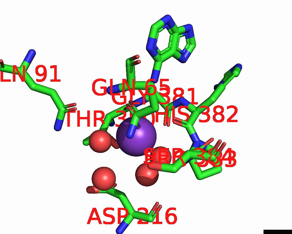
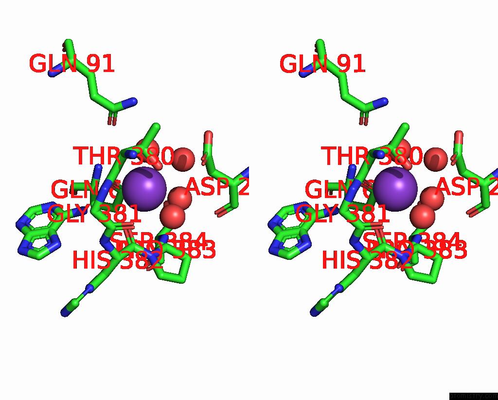
Potassium binding site 1 out of 4 in 8cfv
Go back to
Potassium binding site 1 out
of 4 in the Crystal Structure of S-Adenosyl-L-Homocysteine Hydrolase From P. Aeruginosa in Complex with F2X-Entry Library Fragment H04
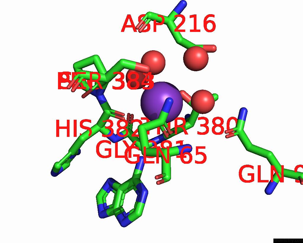
Mono view
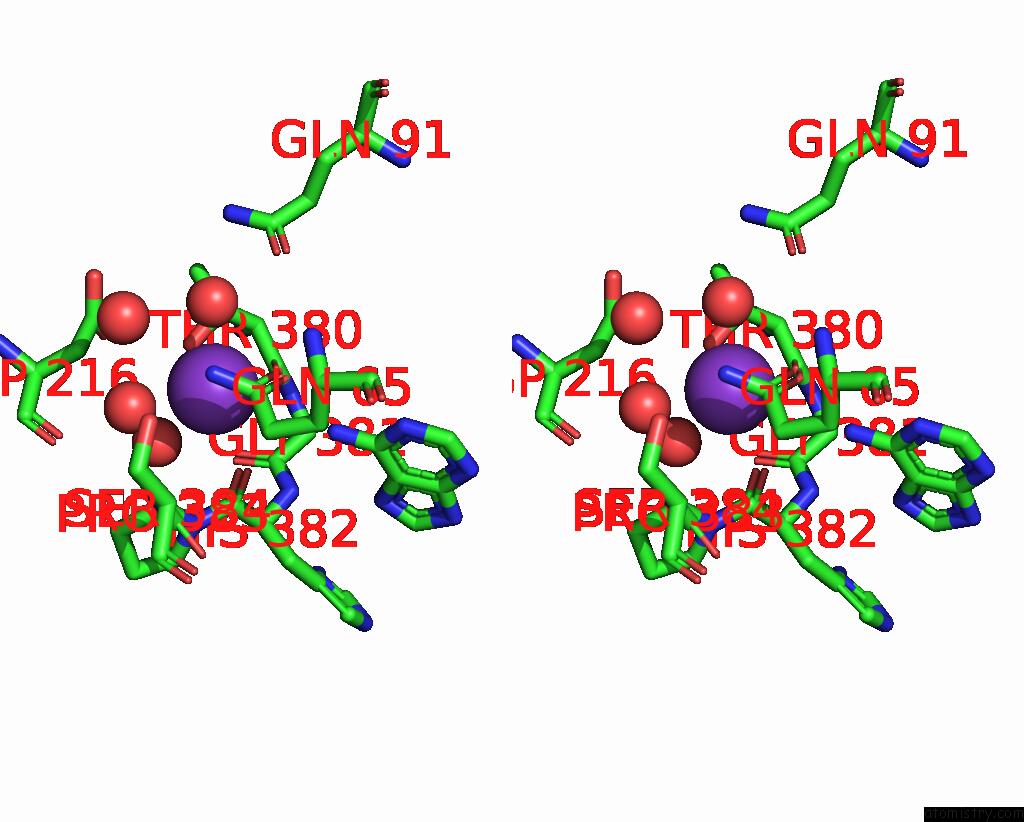
Stereo pair view
Mono view
Stereo pair view
A full contact list of Potassium with other atoms in the K binding
site number 1 of Crystal Structure of S-Adenosyl-L-Homocysteine Hydrolase From P. Aeruginosa in Complex with F2X-Entry Library Fragment H04 within 5.0Å range:
|
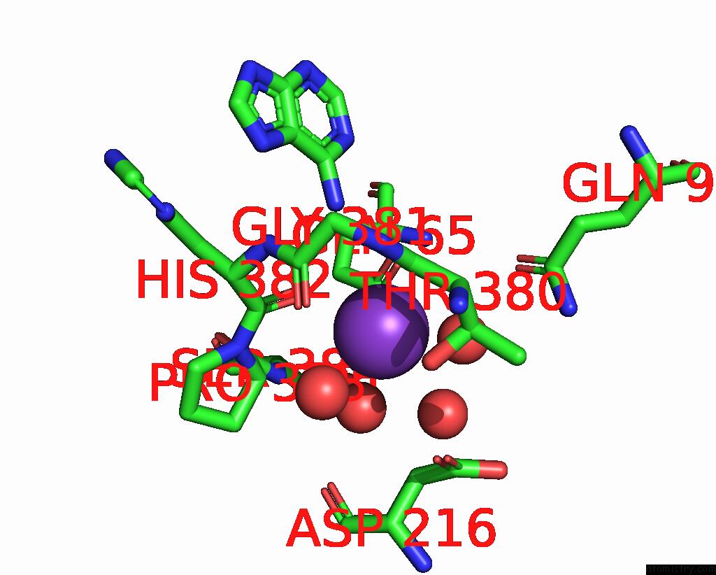
Potassium binding site 2 out of 4 in 8cfv
Go back to
Potassium binding site 2 out
of 4 in the Crystal Structure of S-Adenosyl-L-Homocysteine Hydrolase From P. Aeruginosa in Complex with F2X-Entry Library Fragment H04
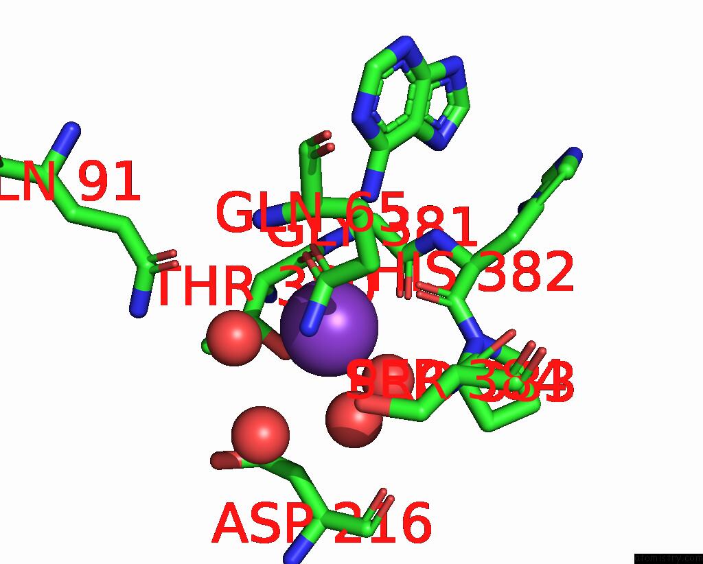
Mono view
Stereo pair view
Mono view
Stereo pair view
A full contact list of Potassium with other atoms in the K binding
site number 2 of Crystal Structure of S-Adenosyl-L-Homocysteine Hydrolase From P. Aeruginosa in Complex with F2X-Entry Library Fragment H04 within 5.0Å range:
|
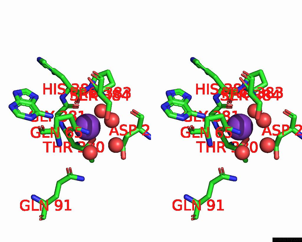
Potassium binding site 3 out of 4 in 8cfv
Go back to
Potassium binding site 3 out
of 4 in the Crystal Structure of S-Adenosyl-L-Homocysteine Hydrolase From P. Aeruginosa in Complex with F2X-Entry Library Fragment H04
Mono view

Stereo pair view
Mono view
Stereo pair view
A full contact list of Potassium with other atoms in the K binding
site number 3 of Crystal Structure of S-Adenosyl-L-Homocysteine Hydrolase From P. Aeruginosa in Complex with F2X-Entry Library Fragment H04 within 5.0Å range:
|
Potassium binding site 4 out of 4 in 8cfv
Go back to
Potassium binding site 4 out
of 4 in the Crystal Structure of S-Adenosyl-L-Homocysteine Hydrolase From P. Aeruginosa in Complex with F2X-Entry Library Fragment H04
Mono view
Stereo pair view
Mono view
Stereo pair view
A full contact list of Potassium with other atoms in the K binding
site number 4 of Crystal Structure of S-Adenosyl-L-Homocysteine Hydrolase From P. Aeruginosa in Complex with F2X-Entry Library Fragment H04 within 5.0Å range:
|
Reference:
P.H.Malecki,
M.Gawel,
M.Stepniewska,
K.Brzezinski.
Crystal Structure of S-Adenosyl-L-Homocysteine Hydrolase From P. Aeruginosa in Complex with Fragment F2X-Entry H04 To Be Published.
Page generated: Sat Aug 9 16:04:18 2025
Last articles
Mg in 3SE5Mg in 3SDK
Mg in 3SDV
Mg in 3SDU
Mg in 3SDT
Mg in 3SDR
Mg in 3SBF
Mg in 3SB5
Mg in 3SCY
Mg in 3SBE