Potassium »
PDB 8b31-8cfg »
8bva »
Potassium in PDB 8bva: Crystal Structure of Mus Musculus Protein Arginine Methyltransferase 2 in Complex with RSF1_114-126
Protein crystallography data
The structure of Crystal Structure of Mus Musculus Protein Arginine Methyltransferase 2 in Complex with RSF1_114-126, PDB code: 8bva
was solved by
V.Cura,
N.Troffer-Charlier,
N.Marechal,
L.Bonnefond,
J.Cavarelli,
with X-Ray Crystallography technique. A brief refinement statistics is given in the table below:
| Resolution Low / High (Å) | 29.55 / 2.19 |
| Space group | C 2 2 21 |
| Cell size a, b, c (Å), α, β, γ (°) | 65.711, 114.409, 133.065, 90, 90, 90 |
| R / Rfree (%) | 17.7 / 20.2 |
Potassium Binding Sites:
The binding sites of Potassium atom in the Crystal Structure of Mus Musculus Protein Arginine Methyltransferase 2 in Complex with RSF1_114-126
(pdb code 8bva). This binding sites where shown within
5.0 Angstroms radius around Potassium atom.
In total only one binding site of Potassium was determined in the Crystal Structure of Mus Musculus Protein Arginine Methyltransferase 2 in Complex with RSF1_114-126, PDB code: 8bva:
In total only one binding site of Potassium was determined in the Crystal Structure of Mus Musculus Protein Arginine Methyltransferase 2 in Complex with RSF1_114-126, PDB code: 8bva:
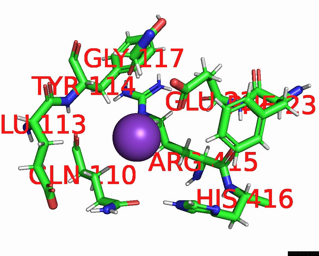
Potassium binding site 1 out of 1 in 8bva
Go back to
Potassium binding site 1 out
of 1 in the Crystal Structure of Mus Musculus Protein Arginine Methyltransferase 2 in Complex with RSF1_114-126
Mono view

Stereo pair view
Mono view
Stereo pair view
A full contact list of Potassium with other atoms in the K binding
site number 1 of Crystal Structure of Mus Musculus Protein Arginine Methyltransferase 2 in Complex with RSF1_114-126 within 5.0Å range:
|
Reference:
V.Cura,
N.Troffer-Charlier,
N.Marechal,
L.Bonnefond,
J.Cavarelli.
Crystal Structure of Mus Musculus Protein Arginine Methyltransferase 2 in Complex with RSF1_114-126 To Be Published.
Page generated: Sat Aug 9 15:40:30 2025
Last articles
Mg in 5WNVMg in 5WNT
Mg in 5WU3
Mg in 5WTI
Mg in 5WSK
Mg in 5WSG
Mg in 5WSC
Mg in 5WSB
Mg in 5WSA
Mg in 5WS9