Potassium »
PDB 7zri-8bm0 »
8azf »
Potassium in PDB 8azf: Crystal Structure of K449E Variant of S-Adenosyl-L-Homocysteine Hydrolase From Pseudomonas Aeruginosa in Complex with Adenosine
Enzymatic activity of Crystal Structure of K449E Variant of S-Adenosyl-L-Homocysteine Hydrolase From Pseudomonas Aeruginosa in Complex with Adenosine
All present enzymatic activity of Crystal Structure of K449E Variant of S-Adenosyl-L-Homocysteine Hydrolase From Pseudomonas Aeruginosa in Complex with Adenosine:
3.3.1.1;
3.3.1.1;
Protein crystallography data
The structure of Crystal Structure of K449E Variant of S-Adenosyl-L-Homocysteine Hydrolase From Pseudomonas Aeruginosa in Complex with Adenosine, PDB code: 8azf
was solved by
A.Arning,
P.Malecki,
K.Wozniak,
K.Brzezinski,
with X-Ray Crystallography technique. A brief refinement statistics is given in the table below:
| Resolution Low / High (Å) | 67.13 / 1.41 |
| Space group | C 1 2 1 |
| Cell size a, b, c (Å), α, β, γ (°) | 142.881, 85.916, 111.824, 90, 122.08, 90 |
| R / Rfree (%) | 16.7 / 22.5 |
Potassium Binding Sites:
The binding sites of Potassium atom in the Crystal Structure of K449E Variant of S-Adenosyl-L-Homocysteine Hydrolase From Pseudomonas Aeruginosa in Complex with Adenosine
(pdb code 8azf). This binding sites where shown within
5.0 Angstroms radius around Potassium atom.
In total 2 binding sites of Potassium where determined in the Crystal Structure of K449E Variant of S-Adenosyl-L-Homocysteine Hydrolase From Pseudomonas Aeruginosa in Complex with Adenosine, PDB code: 8azf:
Jump to Potassium binding site number: 1; 2;
In total 2 binding sites of Potassium where determined in the Crystal Structure of K449E Variant of S-Adenosyl-L-Homocysteine Hydrolase From Pseudomonas Aeruginosa in Complex with Adenosine, PDB code: 8azf:
Jump to Potassium binding site number: 1; 2;
Potassium binding site 1 out of 2 in 8azf
Go back to
Potassium binding site 1 out
of 2 in the Crystal Structure of K449E Variant of S-Adenosyl-L-Homocysteine Hydrolase From Pseudomonas Aeruginosa in Complex with Adenosine
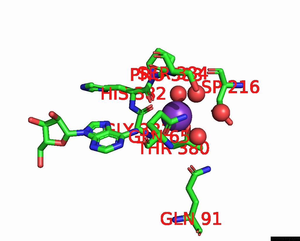
Mono view
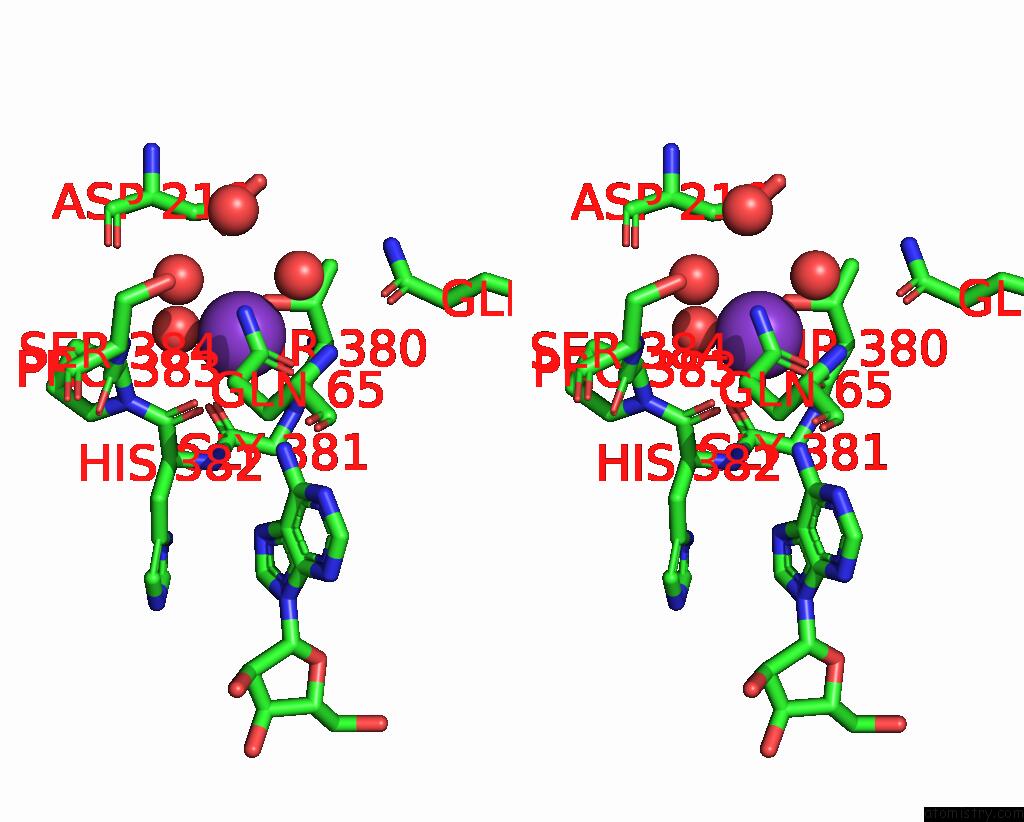
Stereo pair view
Mono view
Stereo pair view
A full contact list of Potassium with other atoms in the K binding
site number 1 of Crystal Structure of K449E Variant of S-Adenosyl-L-Homocysteine Hydrolase From Pseudomonas Aeruginosa in Complex with Adenosine within 5.0Å range:
|
Potassium binding site 2 out of 2 in 8azf
Go back to
Potassium binding site 2 out
of 2 in the Crystal Structure of K449E Variant of S-Adenosyl-L-Homocysteine Hydrolase From Pseudomonas Aeruginosa in Complex with Adenosine
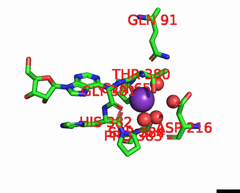
Mono view

Stereo pair view
Mono view
Stereo pair view
A full contact list of Potassium with other atoms in the K binding
site number 2 of Crystal Structure of K449E Variant of S-Adenosyl-L-Homocysteine Hydrolase From Pseudomonas Aeruginosa in Complex with Adenosine within 5.0Å range:
|
Reference:
A.Arning,
P.Malecki,
K.Wozniak,
K.Brzezinski.
Crystal Structure of K449E Variant of S-Adenosyl-L-Homocysteine Hydrolase From Pseudomonas Aeruginosa in Complex with Adenosine To Be Published.
Page generated: Mon Aug 12 22:01:15 2024
Last articles
Zn in 9JPJZn in 9JP7
Zn in 9JPK
Zn in 9JPL
Zn in 9GN6
Zn in 9GN7
Zn in 9GKU
Zn in 9GKW
Zn in 9GKX
Zn in 9GL0