Potassium »
PDB 7zri-8bm0 »
8a71 »
Potassium in PDB 8a71: Crystal Structure of Right-Handed Z-Dna Containing 2'-Deoxy-L-Ribose in Complex with the Polyamine Cadaverine and Potassium Cations at Ultrahigh Resolution
Protein crystallography data
The structure of Crystal Structure of Right-Handed Z-Dna Containing 2'-Deoxy-L-Ribose in Complex with the Polyamine Cadaverine and Potassium Cations at Ultrahigh Resolution, PDB code: 8a71
was solved by
P.Drozdzal,
T.Manszewski,
M.Gilski,
K.Brzezinski,
M.Jaskolski,
with X-Ray Crystallography technique. A brief refinement statistics is given in the table below:
| Resolution Low / High (Å) | 25.43 / 0.69 |
| Space group | P 21 21 21 |
| Cell size a, b, c (Å), α, β, γ (°) | 17.914, 31.127, 44.106, 90, 90, 90 |
| R / Rfree (%) | n/a / n/a |
Potassium Binding Sites:
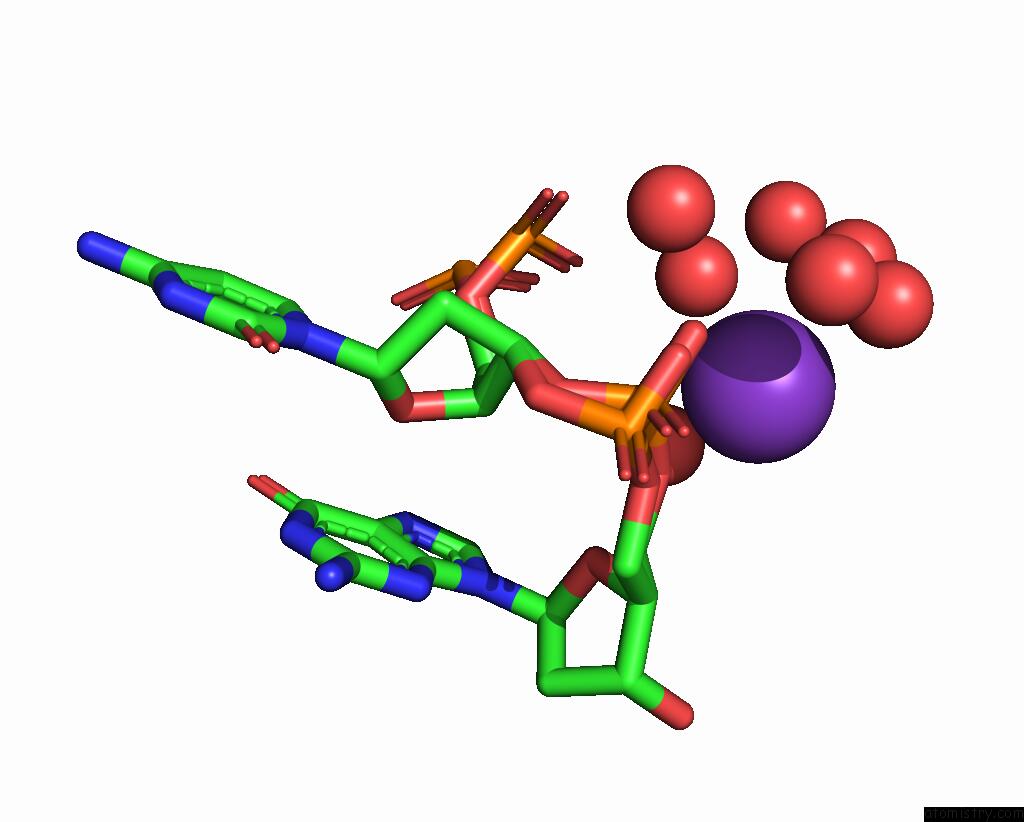
The binding sites of Potassium atom in the Crystal Structure of Right-Handed Z-Dna Containing 2'-Deoxy-L-Ribose in Complex with the Polyamine Cadaverine and Potassium Cations at Ultrahigh Resolution
(pdb code 8a71). This binding sites where shown within
5.0 Angstroms radius around Potassium atom.
In total only one binding site of Potassium was determined in the Crystal Structure of Right-Handed Z-Dna Containing 2'-Deoxy-L-Ribose in Complex with the Polyamine Cadaverine and Potassium Cations at Ultrahigh Resolution, PDB code: 8a71:
In total only one binding site of Potassium was determined in the Crystal Structure of Right-Handed Z-Dna Containing 2'-Deoxy-L-Ribose in Complex with the Polyamine Cadaverine and Potassium Cations at Ultrahigh Resolution, PDB code: 8a71:
Potassium binding site 1 out of 1 in 8a71
Go back to
Potassium binding site 1 out
of 1 in the Crystal Structure of Right-Handed Z-Dna Containing 2'-Deoxy-L-Ribose in Complex with the Polyamine Cadaverine and Potassium Cations at Ultrahigh Resolution
Mono view

Stereo pair view
Mono view
Stereo pair view
A full contact list of Potassium with other atoms in the K binding
site number 1 of Crystal Structure of Right-Handed Z-Dna Containing 2'-Deoxy-L-Ribose in Complex with the Polyamine Cadaverine and Potassium Cations at Ultrahigh Resolution within 5.0Å range:
|
Reference:
P.Drozdzal,
T.Manszewski,
M.Gilski,
K.Brzezinski,
M.Jaskolski.
Right-Handed Z-Dna at Ultrahigh Resolution: A Tale of Two Hands and the Power of the Crystallographic Method. Acta Crystallogr D Struct V. 79 133 2023BIOL.
ISSN: ISSN 2059-7983
PubMed: 36762859
DOI: 10.1107/S2059798322011937
Page generated: Mon Aug 12 21:59:30 2024
ISSN: ISSN 2059-7983
PubMed: 36762859
DOI: 10.1107/S2059798322011937
Last articles
Zn in 9JPJZn in 9JP7
Zn in 9JPK
Zn in 9JPL
Zn in 9GN6
Zn in 9GN7
Zn in 9GKU
Zn in 9GKW
Zn in 9GKX
Zn in 9GL0