Potassium »
PDB 7yze-7zrh »
7zkn »
Potassium in PDB 7zkn: X-Ray Structure of the Complex Between Human Alpha Thrombin and A Pseudo-Cyclic Thrombin Binding Aptamer (Tba-Nnp/Ddp) - Crystal Form Gamma
Enzymatic activity of X-Ray Structure of the Complex Between Human Alpha Thrombin and A Pseudo-Cyclic Thrombin Binding Aptamer (Tba-Nnp/Ddp) - Crystal Form Gamma
All present enzymatic activity of X-Ray Structure of the Complex Between Human Alpha Thrombin and A Pseudo-Cyclic Thrombin Binding Aptamer (Tba-Nnp/Ddp) - Crystal Form Gamma:
3.4.21.5;
3.4.21.5;
Protein crystallography data
The structure of X-Ray Structure of the Complex Between Human Alpha Thrombin and A Pseudo-Cyclic Thrombin Binding Aptamer (Tba-Nnp/Ddp) - Crystal Form Gamma, PDB code: 7zkn
was solved by
R.Troisi,
F.Sica,
with X-Ray Crystallography technique. A brief refinement statistics is given in the table below:
| Resolution Low / High (Å) | 68.13 / 3.03 |
| Space group | P 1 21 1 |
| Cell size a, b, c (Å), α, β, γ (°) | 76.62, 114.86, 83.44, 90, 117.25, 90 |
| R / Rfree (%) | 20.1 / 24.3 |
Potassium Binding Sites:
The binding sites of Potassium atom in the X-Ray Structure of the Complex Between Human Alpha Thrombin and A Pseudo-Cyclic Thrombin Binding Aptamer (Tba-Nnp/Ddp) - Crystal Form Gamma
(pdb code 7zkn). This binding sites where shown within
5.0 Angstroms radius around Potassium atom.
In total 4 binding sites of Potassium where determined in the X-Ray Structure of the Complex Between Human Alpha Thrombin and A Pseudo-Cyclic Thrombin Binding Aptamer (Tba-Nnp/Ddp) - Crystal Form Gamma, PDB code: 7zkn:
Jump to Potassium binding site number: 1; 2; 3; 4;
In total 4 binding sites of Potassium where determined in the X-Ray Structure of the Complex Between Human Alpha Thrombin and A Pseudo-Cyclic Thrombin Binding Aptamer (Tba-Nnp/Ddp) - Crystal Form Gamma, PDB code: 7zkn:
Jump to Potassium binding site number: 1; 2; 3; 4;
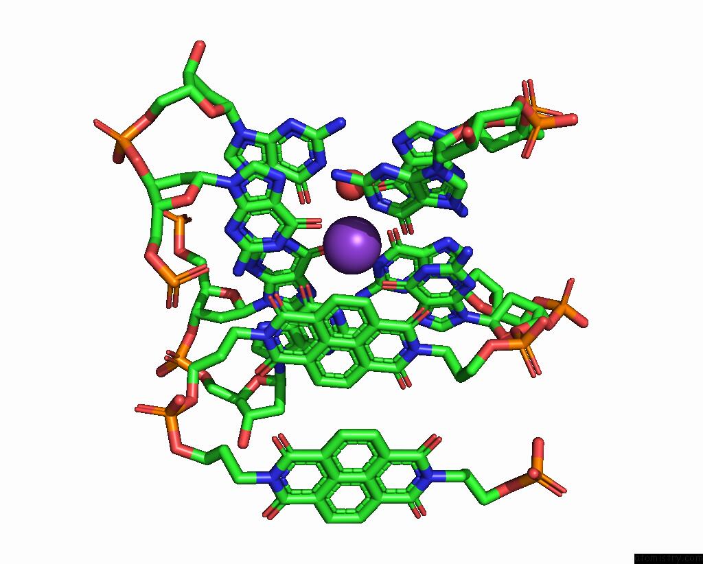
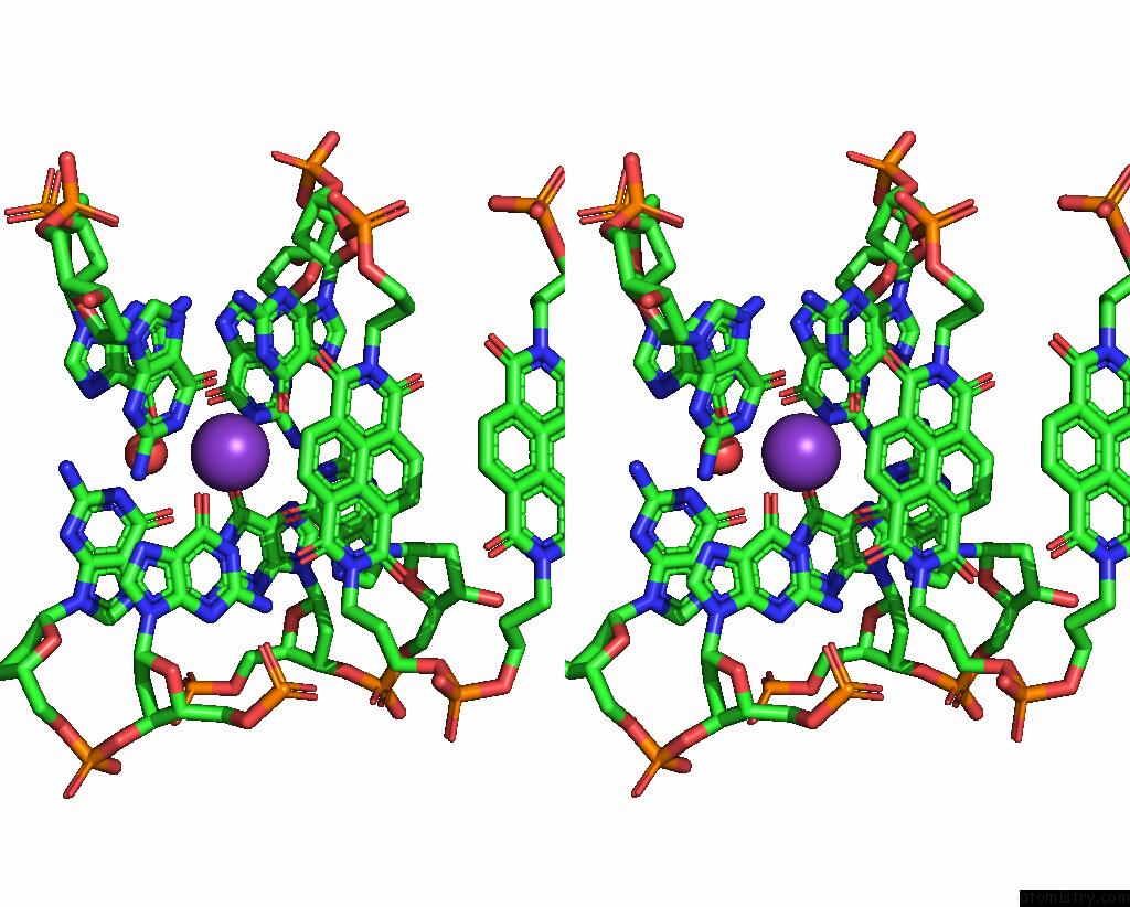
Potassium binding site 1 out of 4 in 7zkn
Go back to
Potassium binding site 1 out
of 4 in the X-Ray Structure of the Complex Between Human Alpha Thrombin and A Pseudo-Cyclic Thrombin Binding Aptamer (Tba-Nnp/Ddp) - Crystal Form Gamma
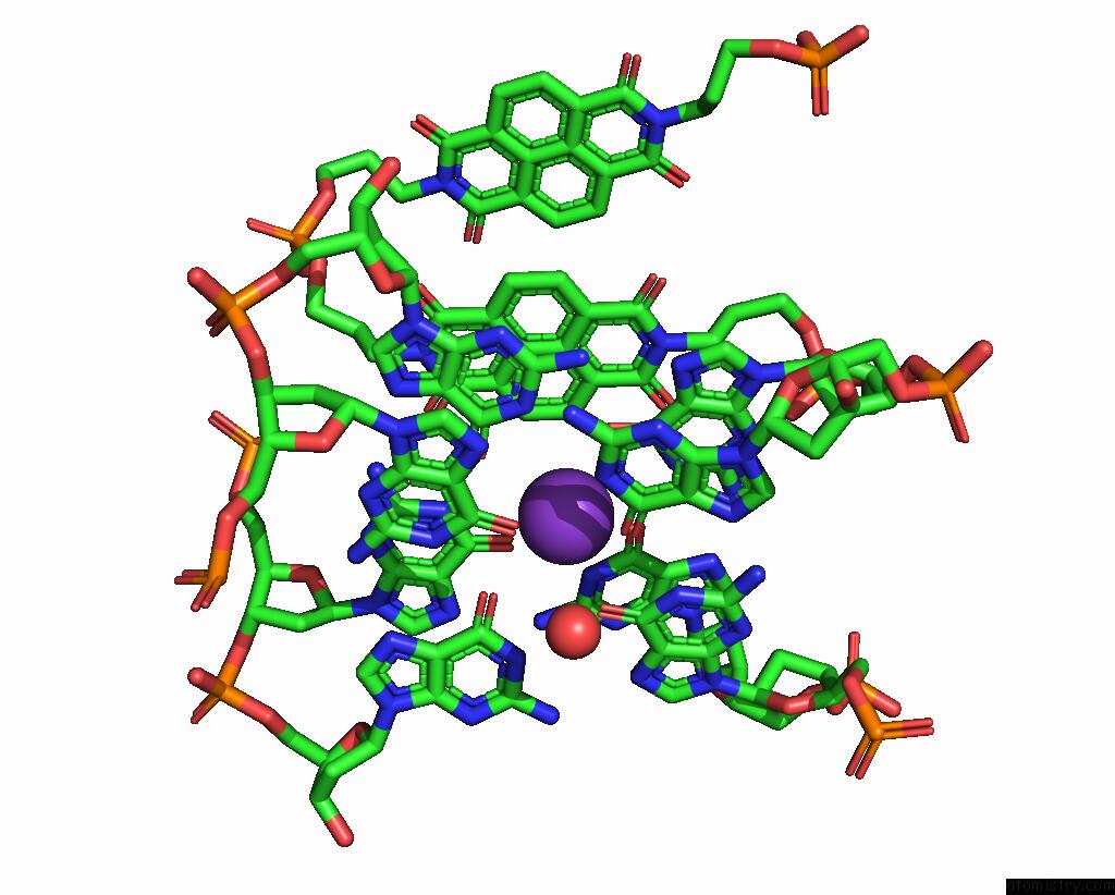
Mono view
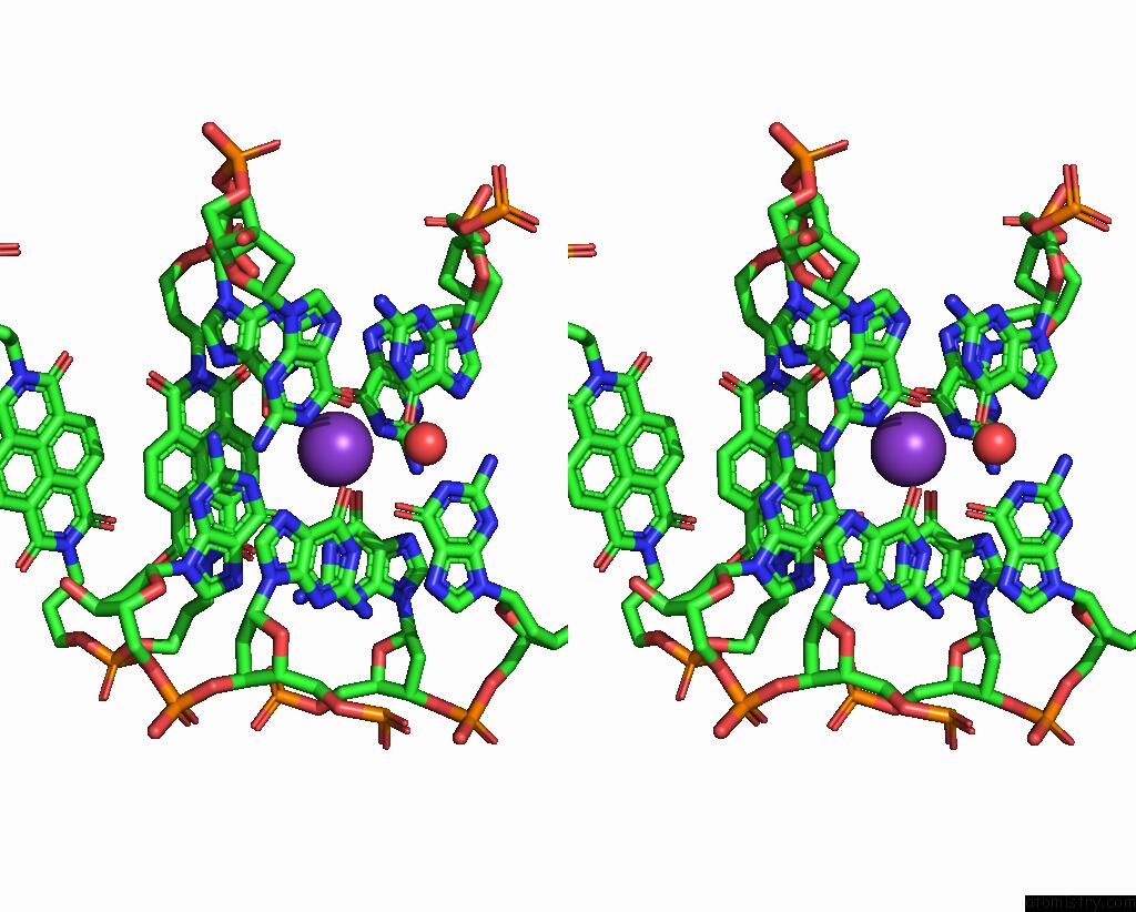
Stereo pair view
Mono view
Stereo pair view
A full contact list of Potassium with other atoms in the K binding
site number 1 of X-Ray Structure of the Complex Between Human Alpha Thrombin and A Pseudo-Cyclic Thrombin Binding Aptamer (Tba-Nnp/Ddp) - Crystal Form Gamma within 5.0Å range:
|
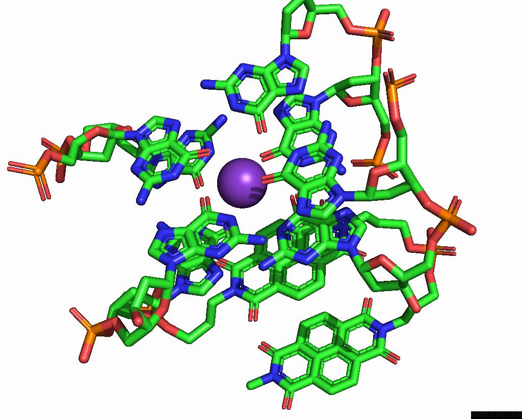
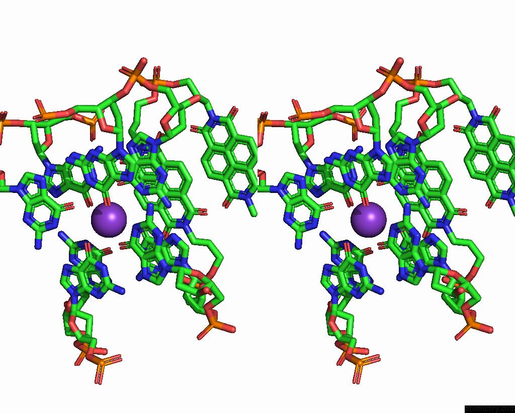
Potassium binding site 2 out of 4 in 7zkn
Go back to
Potassium binding site 2 out
of 4 in the X-Ray Structure of the Complex Between Human Alpha Thrombin and A Pseudo-Cyclic Thrombin Binding Aptamer (Tba-Nnp/Ddp) - Crystal Form Gamma
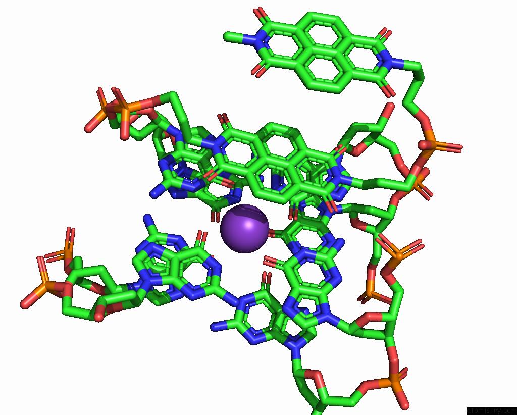
Mono view
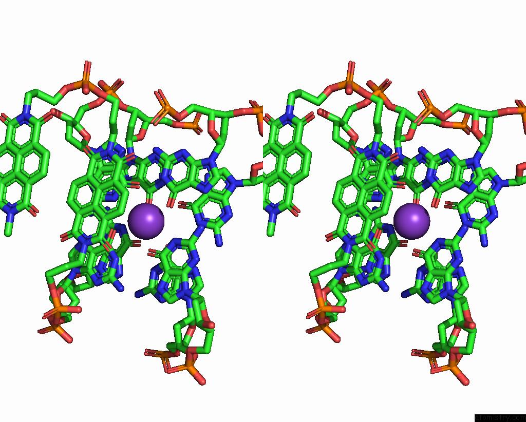
Stereo pair view
Mono view
Stereo pair view
A full contact list of Potassium with other atoms in the K binding
site number 2 of X-Ray Structure of the Complex Between Human Alpha Thrombin and A Pseudo-Cyclic Thrombin Binding Aptamer (Tba-Nnp/Ddp) - Crystal Form Gamma within 5.0Å range:
|
Potassium binding site 3 out of 4 in 7zkn
Go back to
Potassium binding site 3 out
of 4 in the X-Ray Structure of the Complex Between Human Alpha Thrombin and A Pseudo-Cyclic Thrombin Binding Aptamer (Tba-Nnp/Ddp) - Crystal Form Gamma
Mono view
Stereo pair view
Mono view
Stereo pair view
A full contact list of Potassium with other atoms in the K binding
site number 3 of X-Ray Structure of the Complex Between Human Alpha Thrombin and A Pseudo-Cyclic Thrombin Binding Aptamer (Tba-Nnp/Ddp) - Crystal Form Gamma within 5.0Å range:
|
Potassium binding site 4 out of 4 in 7zkn
Go back to
Potassium binding site 4 out
of 4 in the X-Ray Structure of the Complex Between Human Alpha Thrombin and A Pseudo-Cyclic Thrombin Binding Aptamer (Tba-Nnp/Ddp) - Crystal Form Gamma
Mono view
Stereo pair view
Mono view
Stereo pair view
A full contact list of Potassium with other atoms in the K binding
site number 4 of X-Ray Structure of the Complex Between Human Alpha Thrombin and A Pseudo-Cyclic Thrombin Binding Aptamer (Tba-Nnp/Ddp) - Crystal Form Gamma within 5.0Å range:
|
Reference:
R.Troisi,
C.Riccardi,
K.Perez De Carvasal,
M.Smietana,
F.Morvan,
P.Del Vecchio,
D.Montesarchio,
F.Sica.
A Terminal Functionalization Strategy Reveals Unusual Binding Abilities of Anti-Thrombin Anticoagulant Aptamers. Mol Ther Nucleic Acids V. 30 585 2022.
ISSN: ISSN 2162-2531
PubMed: 36457701
DOI: 10.1016/J.OMTN.2022.11.007
Page generated: Mon Aug 12 21:50:12 2024
ISSN: ISSN 2162-2531
PubMed: 36457701
DOI: 10.1016/J.OMTN.2022.11.007
Last articles
Zn in 9JYWZn in 9IR4
Zn in 9IR3
Zn in 9GMX
Zn in 9GMW
Zn in 9JEJ
Zn in 9ERF
Zn in 9ERE
Zn in 9EGV
Zn in 9EGW