Potassium »
PDB 7xxk-7zgo »
7z3v »
Potassium in PDB 7z3v: Escherichia Coli Periplasmic Phytase Appa D304E Mutant, Complex with Myo-Inositol Hexakissulfate
Enzymatic activity of Escherichia Coli Periplasmic Phytase Appa D304E Mutant, Complex with Myo-Inositol Hexakissulfate
All present enzymatic activity of Escherichia Coli Periplasmic Phytase Appa D304E Mutant, Complex with Myo-Inositol Hexakissulfate:
3.1.3.2; 3.1.3.26;
3.1.3.2; 3.1.3.26;
Protein crystallography data
The structure of Escherichia Coli Periplasmic Phytase Appa D304E Mutant, Complex with Myo-Inositol Hexakissulfate, PDB code: 7z3v
was solved by
I.M.Acquistapace,
C.A.Brearley,
A.M.Hemmings,
with X-Ray Crystallography technique. A brief refinement statistics is given in the table below:
| Resolution Low / High (Å) | 65.28 / 2.60 |
| Space group | P 1 21 1 |
| Cell size a, b, c (Å), α, β, γ (°) | 63.712, 44.35, 66.559, 90, 101.27, 90 |
| R / Rfree (%) | 19.5 / 27.1 |
Potassium Binding Sites:
The binding sites of Potassium atom in the Escherichia Coli Periplasmic Phytase Appa D304E Mutant, Complex with Myo-Inositol Hexakissulfate
(pdb code 7z3v). This binding sites where shown within
5.0 Angstroms radius around Potassium atom.
In total only one binding site of Potassium was determined in the Escherichia Coli Periplasmic Phytase Appa D304E Mutant, Complex with Myo-Inositol Hexakissulfate, PDB code: 7z3v:
In total only one binding site of Potassium was determined in the Escherichia Coli Periplasmic Phytase Appa D304E Mutant, Complex with Myo-Inositol Hexakissulfate, PDB code: 7z3v:
Potassium binding site 1 out of 1 in 7z3v
Go back to
Potassium binding site 1 out
of 1 in the Escherichia Coli Periplasmic Phytase Appa D304E Mutant, Complex with Myo-Inositol Hexakissulfate
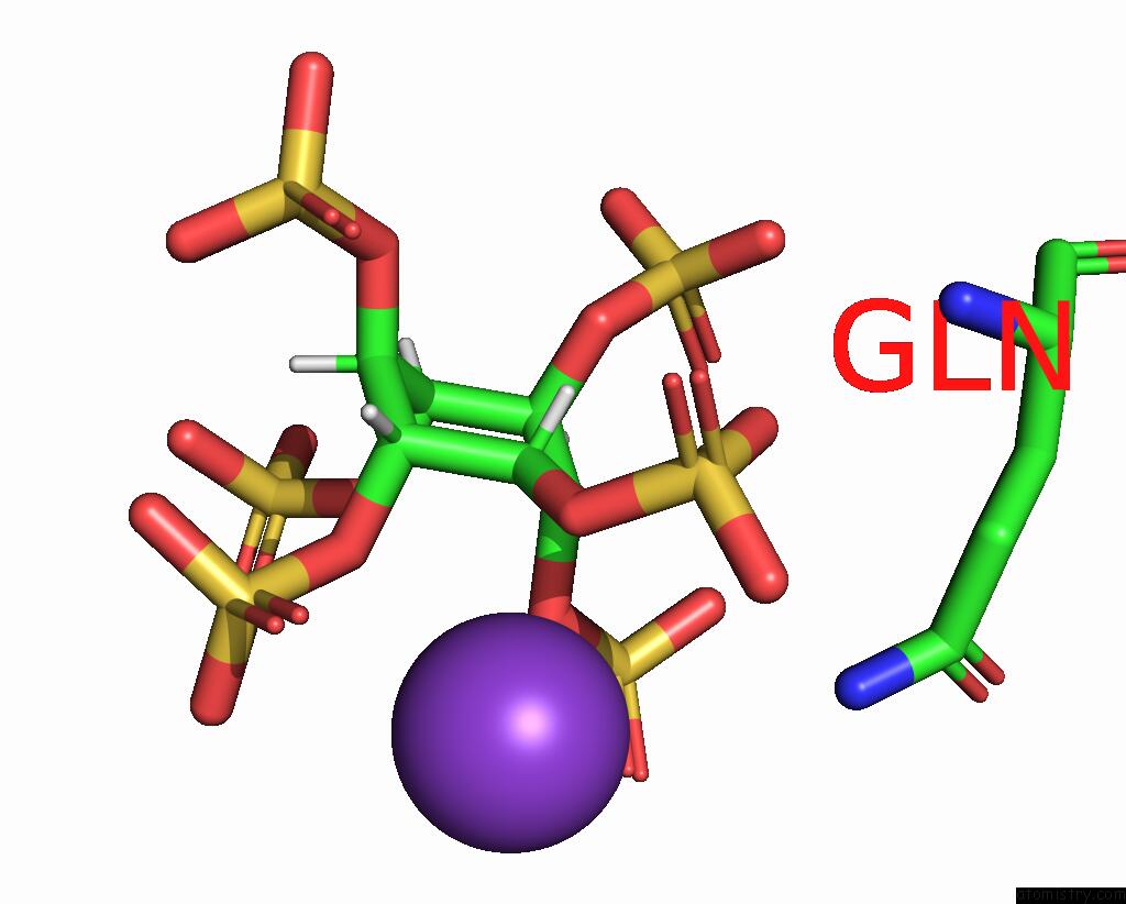
Mono view
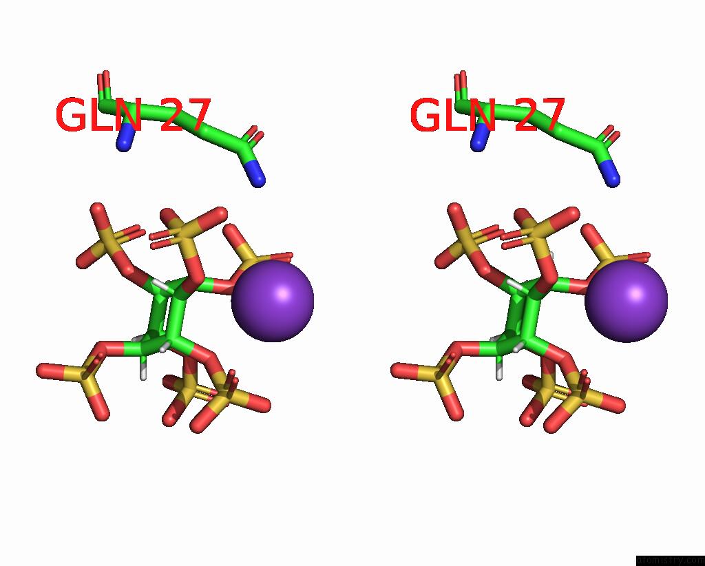
Stereo pair view
Mono view
Stereo pair view
A full contact list of Potassium with other atoms in the K binding
site number 1 of Escherichia Coli Periplasmic Phytase Appa D304E Mutant, Complex with Myo-Inositol Hexakissulfate within 5.0Å range:
|
Reference:
I.M.Acquistapace,
E.J.Thompson,
I.Kuhn,
M.R.Bedford,
C.A.Brearley,
A.M.Hemmings.
Insights to the Structural Basis For the Stereospecificity of the Escherichia Coli Phytase, Appa. Int J Mol Sci V. 23 2022.
ISSN: ESSN 1422-0067
PubMed: 35683026
DOI: 10.3390/IJMS23116346
Page generated: Sat Aug 9 15:21:50 2025
ISSN: ESSN 1422-0067
PubMed: 35683026
DOI: 10.3390/IJMS23116346
Last articles
Mg in 364DMg in 3A0T
Mg in 357D
Mg in 3A06
Mg in 392D
Mg in 389D
Mg in 388D
Mg in 384D
Mg in 354D
Mg in 383D