Potassium »
PDB 7uut-7xx6 »
7xdh »
Potassium in PDB 7xdh: Crystal Structure of A Dimeric Interlocked Parallel G-Quadruplex
Protein crystallography data
The structure of Crystal Structure of A Dimeric Interlocked Parallel G-Quadruplex, PDB code: 7xdh
was solved by
K.H.Ngo,
C.W.Liew,
S.Lattmann,
F.R.Winnerdy,
A.T.Phan,
with X-Ray Crystallography technique. A brief refinement statistics is given in the table below:
| Resolution Low / High (Å) | 29.26 / 1.83 |
| Space group | P 1 |
| Cell size a, b, c (Å), α, β, γ (°) | 25.685, 30.34, 56.493, 79.02, 83.36, 78.2 |
| R / Rfree (%) | 22.7 / 25.3 |
Potassium Binding Sites:
The binding sites of Potassium atom in the Crystal Structure of A Dimeric Interlocked Parallel G-Quadruplex
(pdb code 7xdh). This binding sites where shown within
5.0 Angstroms radius around Potassium atom.
In total 10 binding sites of Potassium where determined in the Crystal Structure of A Dimeric Interlocked Parallel G-Quadruplex, PDB code: 7xdh:
Jump to Potassium binding site number: 1; 2; 3; 4; 5; 6; 7; 8; 9; 10;
In total 10 binding sites of Potassium where determined in the Crystal Structure of A Dimeric Interlocked Parallel G-Quadruplex, PDB code: 7xdh:
Jump to Potassium binding site number: 1; 2; 3; 4; 5; 6; 7; 8; 9; 10;
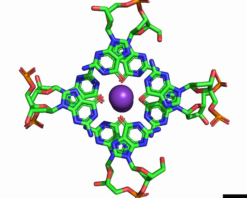
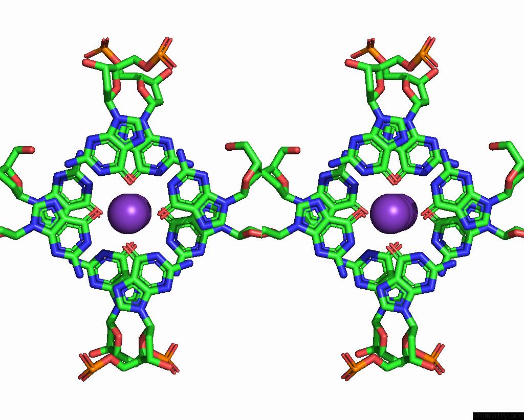
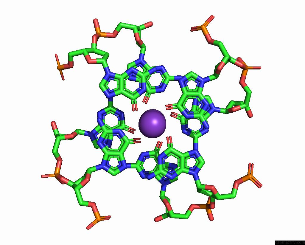
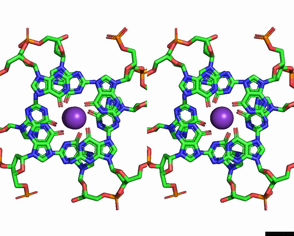
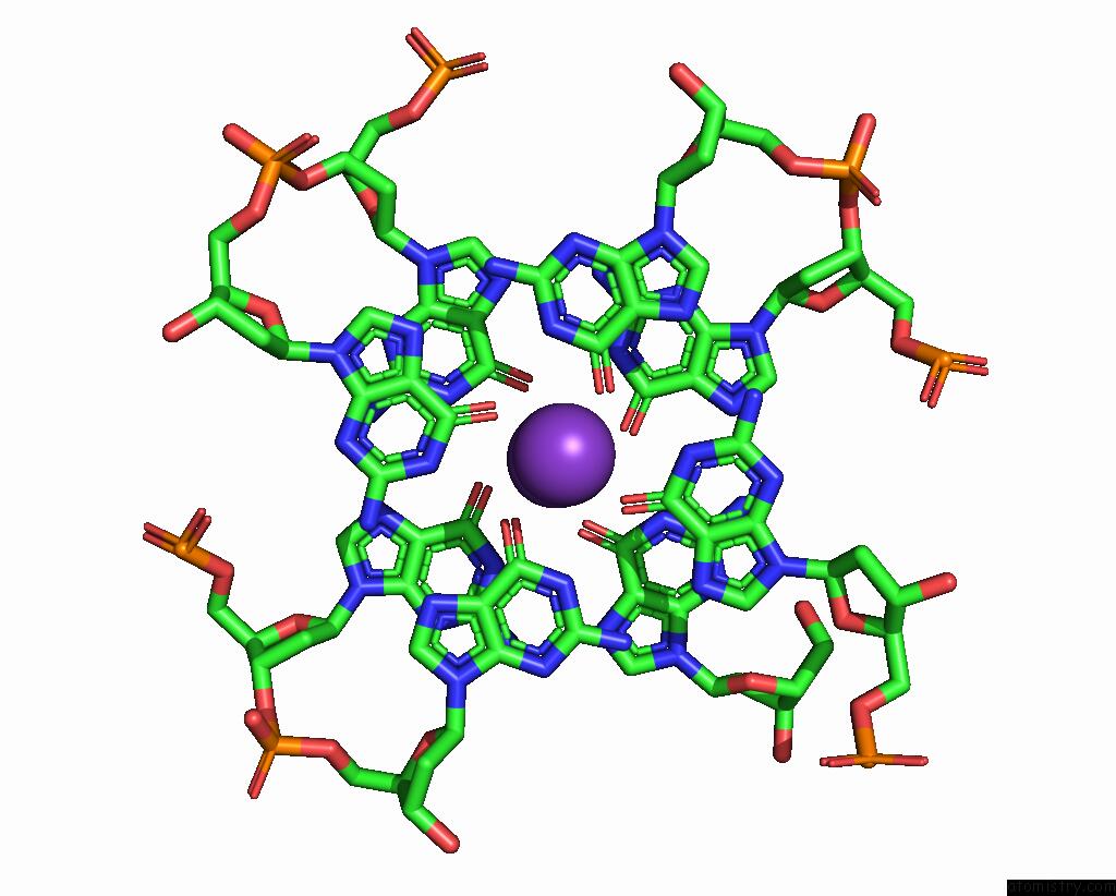
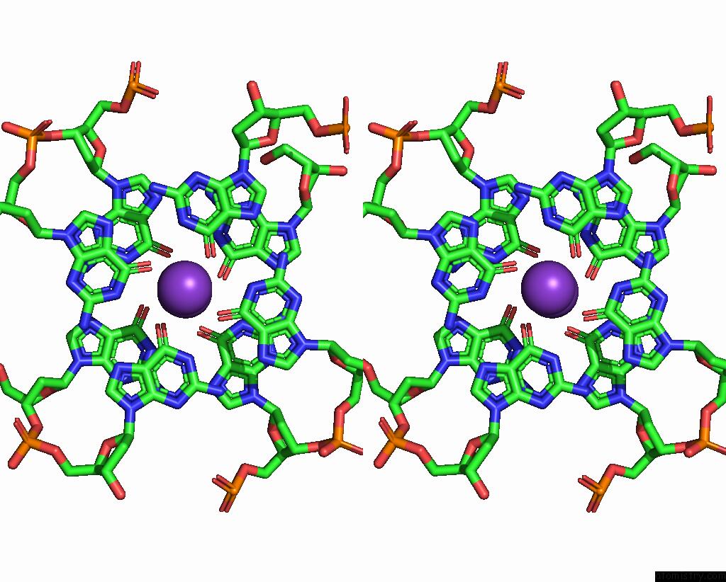
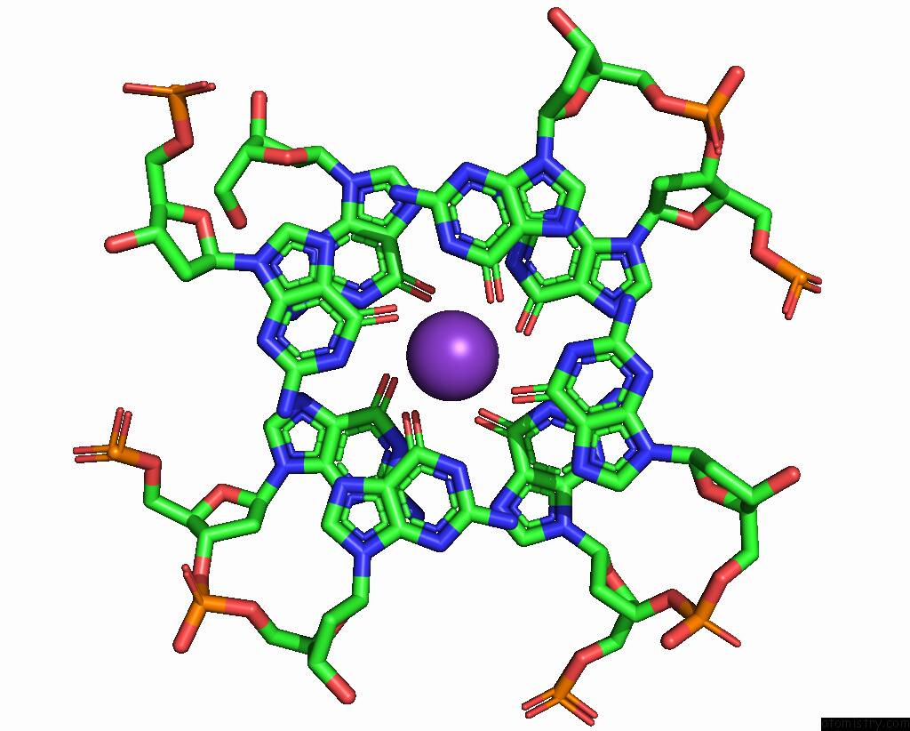
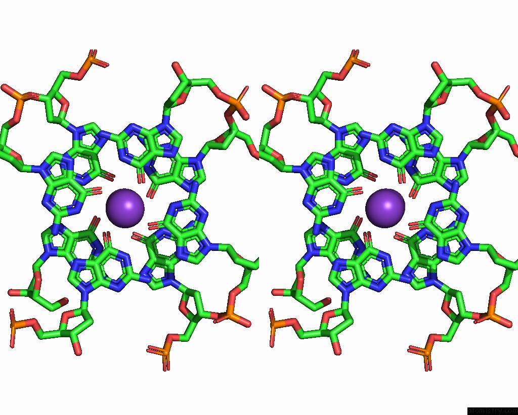
Potassium binding site 1 out of 10 in 7xdh
Go back to
Potassium binding site 1 out
of 10 in the Crystal Structure of A Dimeric Interlocked Parallel G-Quadruplex
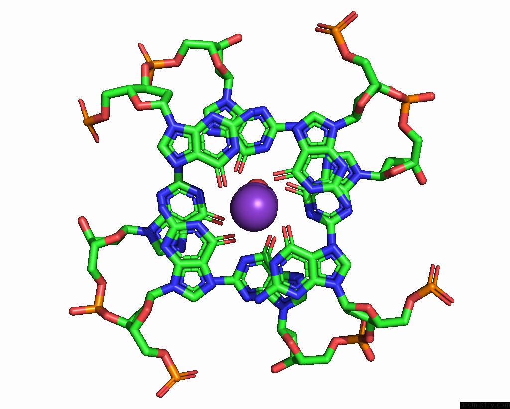
Mono view
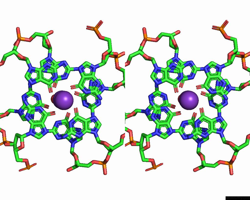
Stereo pair view
Mono view
Stereo pair view
A full contact list of Potassium with other atoms in the K binding
site number 1 of Crystal Structure of A Dimeric Interlocked Parallel G-Quadruplex within 5.0Å range:
|
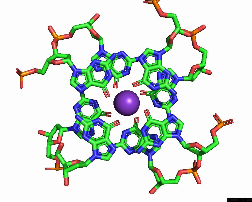
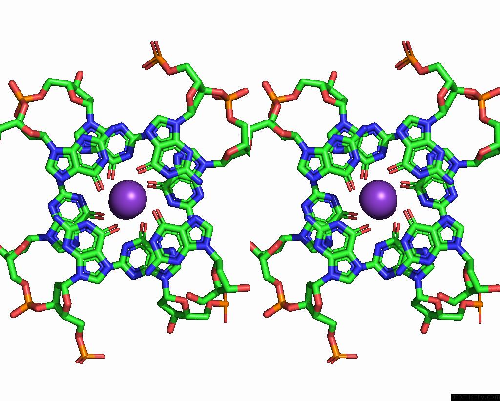
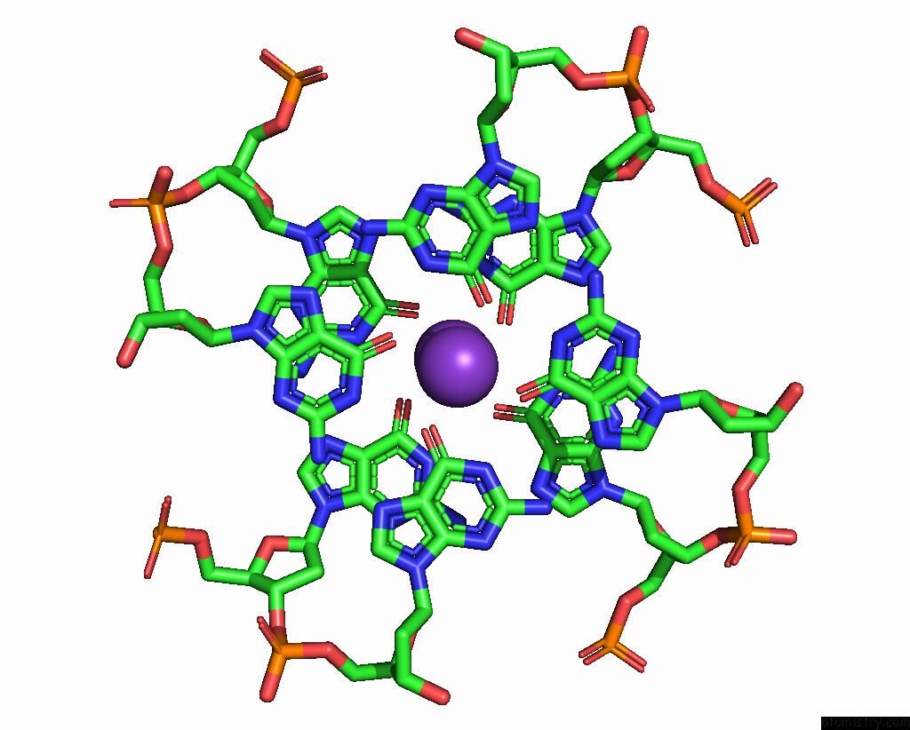
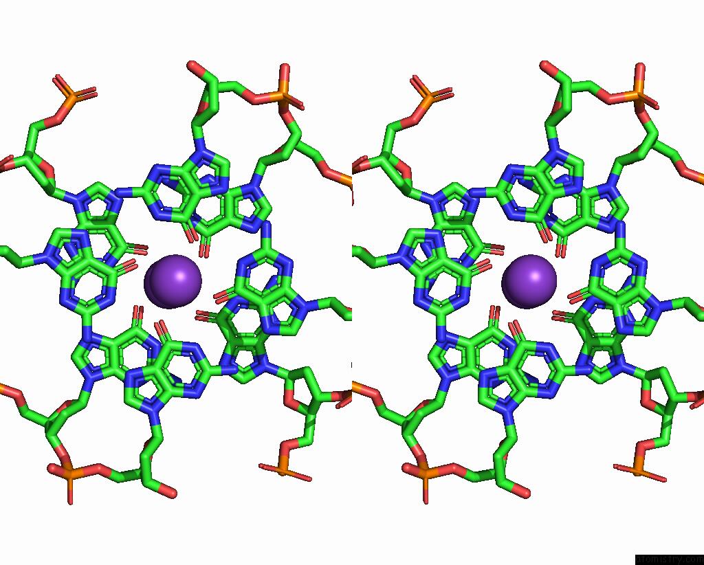
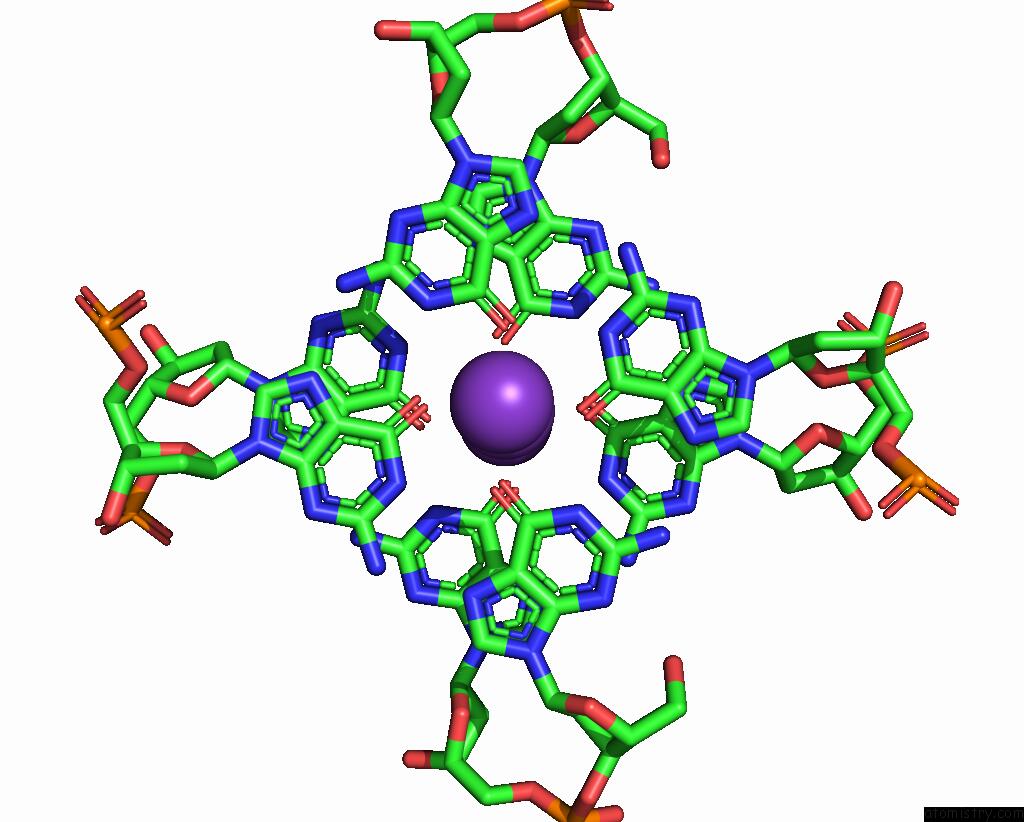
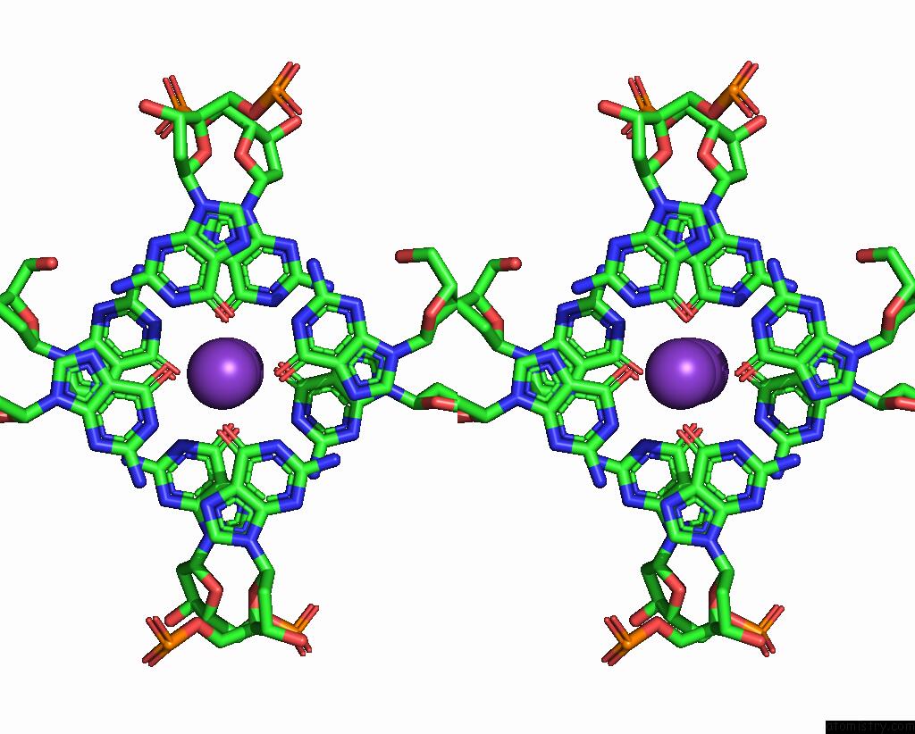
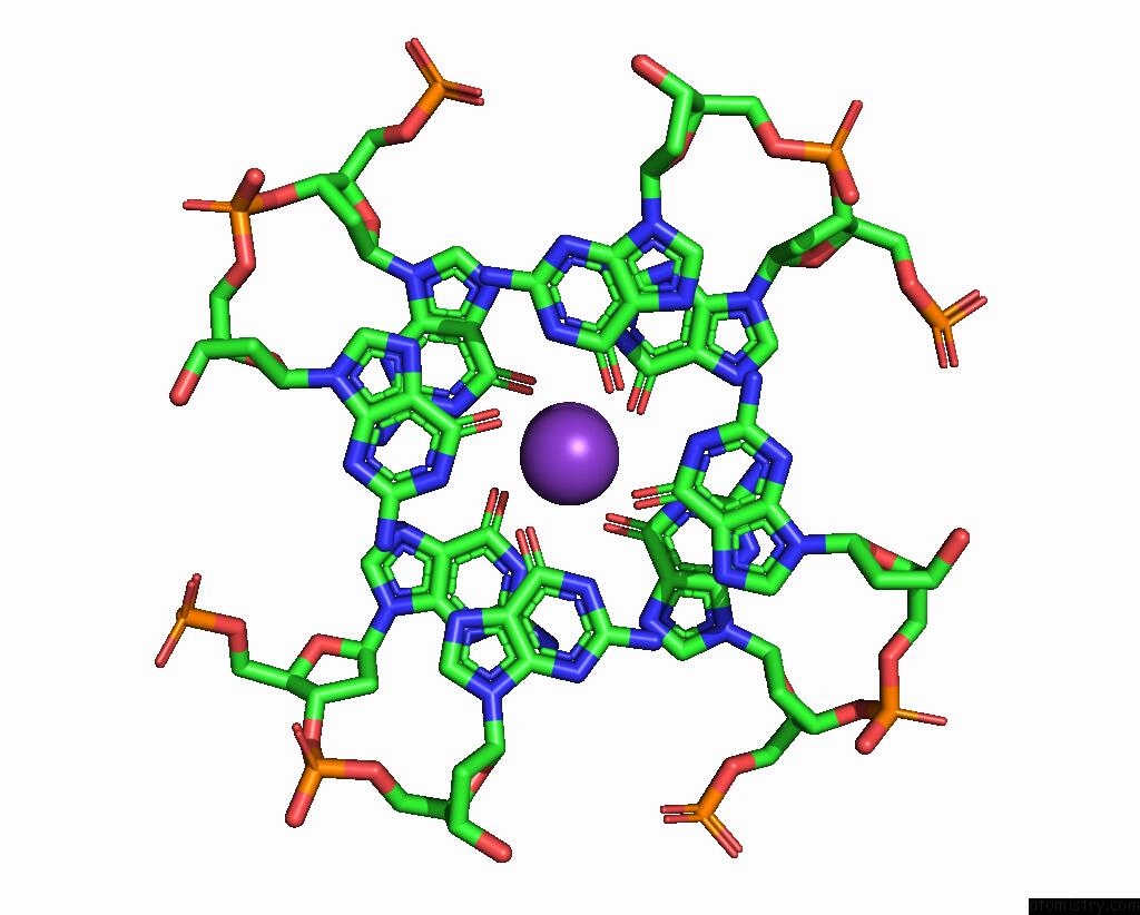
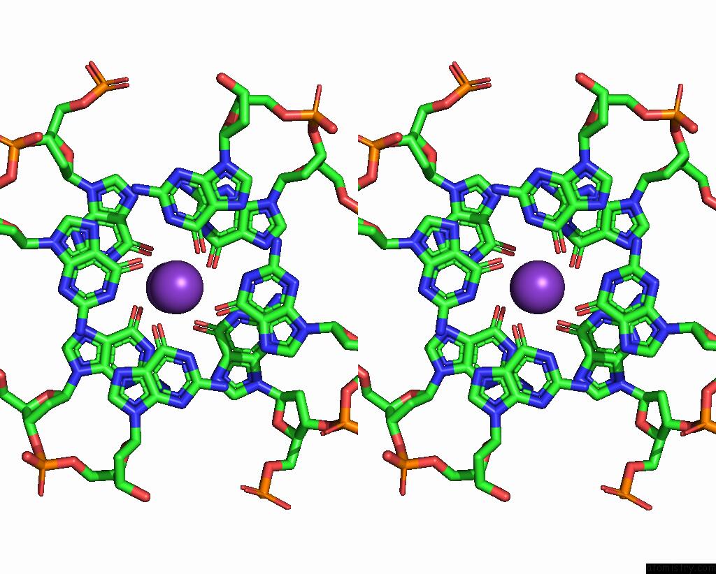
Potassium binding site 2 out of 10 in 7xdh
Go back to
Potassium binding site 2 out
of 10 in the Crystal Structure of A Dimeric Interlocked Parallel G-Quadruplex
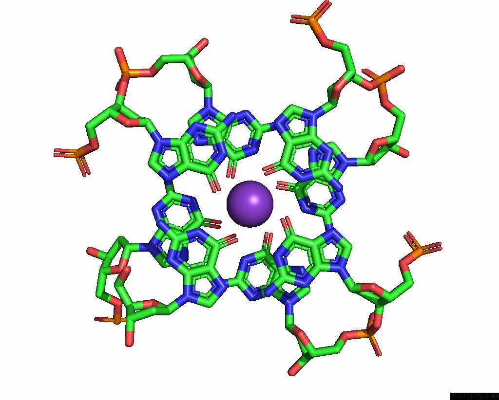
Mono view
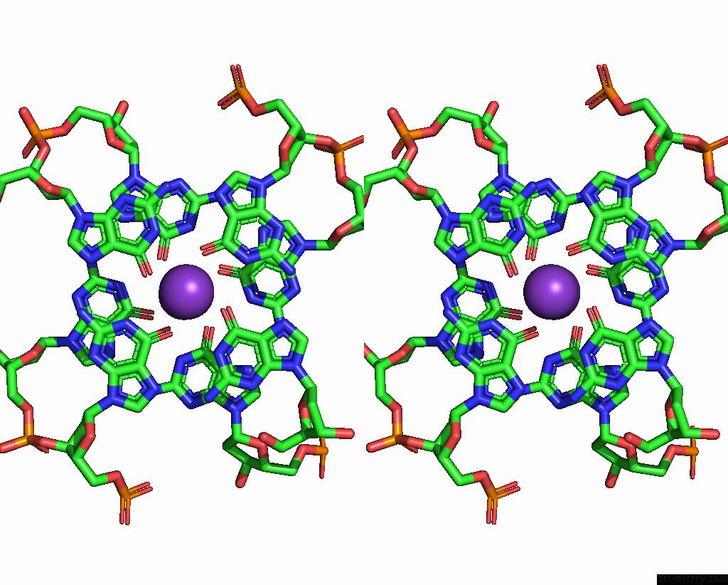
Stereo pair view
Mono view
Stereo pair view
A full contact list of Potassium with other atoms in the K binding
site number 2 of Crystal Structure of A Dimeric Interlocked Parallel G-Quadruplex within 5.0Å range:
|
Potassium binding site 3 out of 10 in 7xdh
Go back to
Potassium binding site 3 out
of 10 in the Crystal Structure of A Dimeric Interlocked Parallel G-Quadruplex
Mono view
Stereo pair view
Mono view
Stereo pair view
A full contact list of Potassium with other atoms in the K binding
site number 3 of Crystal Structure of A Dimeric Interlocked Parallel G-Quadruplex within 5.0Å range:
|
Potassium binding site 4 out of 10 in 7xdh
Go back to
Potassium binding site 4 out
of 10 in the Crystal Structure of A Dimeric Interlocked Parallel G-Quadruplex
Mono view
Stereo pair view
Mono view
Stereo pair view
A full contact list of Potassium with other atoms in the K binding
site number 4 of Crystal Structure of A Dimeric Interlocked Parallel G-Quadruplex within 5.0Å range:
|
Potassium binding site 5 out of 10 in 7xdh
Go back to
Potassium binding site 5 out
of 10 in the Crystal Structure of A Dimeric Interlocked Parallel G-Quadruplex
Mono view
Stereo pair view
Mono view
Stereo pair view
A full contact list of Potassium with other atoms in the K binding
site number 5 of Crystal Structure of A Dimeric Interlocked Parallel G-Quadruplex within 5.0Å range:
|
Potassium binding site 6 out of 10 in 7xdh
Go back to
Potassium binding site 6 out
of 10 in the Crystal Structure of A Dimeric Interlocked Parallel G-Quadruplex
Mono view
Stereo pair view
Mono view
Stereo pair view
A full contact list of Potassium with other atoms in the K binding
site number 6 of Crystal Structure of A Dimeric Interlocked Parallel G-Quadruplex within 5.0Å range:
|
Potassium binding site 7 out of 10 in 7xdh
Go back to
Potassium binding site 7 out
of 10 in the Crystal Structure of A Dimeric Interlocked Parallel G-Quadruplex
Mono view
Stereo pair view
Mono view
Stereo pair view
A full contact list of Potassium with other atoms in the K binding
site number 7 of Crystal Structure of A Dimeric Interlocked Parallel G-Quadruplex within 5.0Å range:
|
Potassium binding site 8 out of 10 in 7xdh
Go back to
Potassium binding site 8 out
of 10 in the Crystal Structure of A Dimeric Interlocked Parallel G-Quadruplex
Mono view
Stereo pair view
Mono view
Stereo pair view
A full contact list of Potassium with other atoms in the K binding
site number 8 of Crystal Structure of A Dimeric Interlocked Parallel G-Quadruplex within 5.0Å range:
|
Potassium binding site 9 out of 10 in 7xdh
Go back to
Potassium binding site 9 out
of 10 in the Crystal Structure of A Dimeric Interlocked Parallel G-Quadruplex
Mono view
Stereo pair view
Mono view
Stereo pair view
A full contact list of Potassium with other atoms in the K binding
site number 9 of Crystal Structure of A Dimeric Interlocked Parallel G-Quadruplex within 5.0Å range:
|
Potassium binding site 10 out of 10 in 7xdh
Go back to
Potassium binding site 10 out
of 10 in the Crystal Structure of A Dimeric Interlocked Parallel G-Quadruplex
Mono view
Stereo pair view
Mono view
Stereo pair view
A full contact list of Potassium with other atoms in the K binding
site number 10 of Crystal Structure of A Dimeric Interlocked Parallel G-Quadruplex within 5.0Å range:
|
Reference:
K.H.Ngo,
C.W.Liew,
S.Lattmann,
F.R.Winnerdy,
A.T.Phan.
Crystal Structures of An Hiv-1 Integrase Aptamer: Formation of A Water-Mediated A.G.G.G.G Pentad in An Interlocked G-Quadruplex Biochem.Biophys.Res.Commun. 2022.
ISSN: ESSN 1090-2104
Page generated: Sat Aug 9 15:12:46 2025
ISSN: ESSN 1090-2104
Last articles
Mg in 3SE5Mg in 3SDK
Mg in 3SDV
Mg in 3SDU
Mg in 3SDT
Mg in 3SDR
Mg in 3SBF
Mg in 3SB5
Mg in 3SCY
Mg in 3SBE