Potassium »
PDB 7uut-7xx6 »
7x7g »
Potassium in PDB 7x7g: Crystal Structure of A Dimeric Interlocked Parallel G-Quadruplex
Protein crystallography data
The structure of Crystal Structure of A Dimeric Interlocked Parallel G-Quadruplex, PDB code: 7x7g
was solved by
K.H.Ngo,
C.W.Liew,
S.Lattmann,
F.R.Winnerdy,
A.T.Phan,
with X-Ray Crystallography technique. A brief refinement statistics is given in the table below:
| Resolution Low / High (Å) | 48.63 / 2.19 |
| Space group | P 1 21 1 |
| Cell size a, b, c (Å), α, β, γ (°) | 26.173, 31.247, 49.893, 90, 102.9, 90 |
| R / Rfree (%) | 25.6 / 26.9 |
Potassium Binding Sites:
The binding sites of Potassium atom in the Crystal Structure of A Dimeric Interlocked Parallel G-Quadruplex
(pdb code 7x7g). This binding sites where shown within
5.0 Angstroms radius around Potassium atom.
In total 5 binding sites of Potassium where determined in the Crystal Structure of A Dimeric Interlocked Parallel G-Quadruplex, PDB code: 7x7g:
Jump to Potassium binding site number: 1; 2; 3; 4; 5;
In total 5 binding sites of Potassium where determined in the Crystal Structure of A Dimeric Interlocked Parallel G-Quadruplex, PDB code: 7x7g:
Jump to Potassium binding site number: 1; 2; 3; 4; 5;
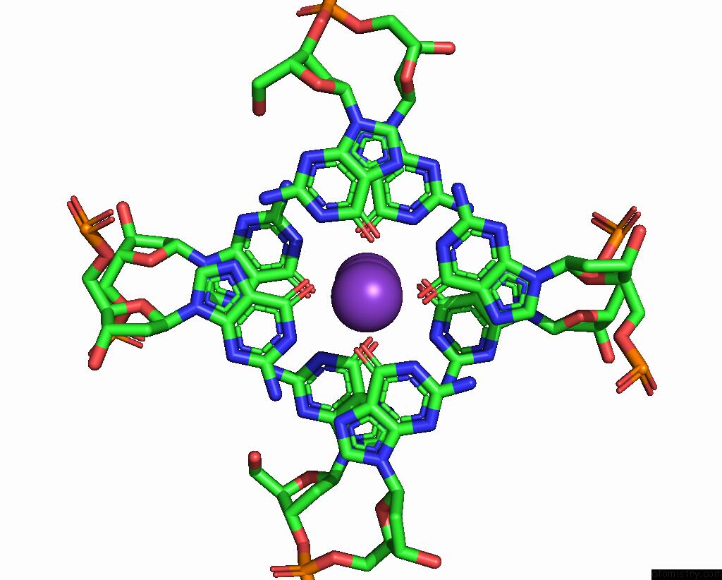
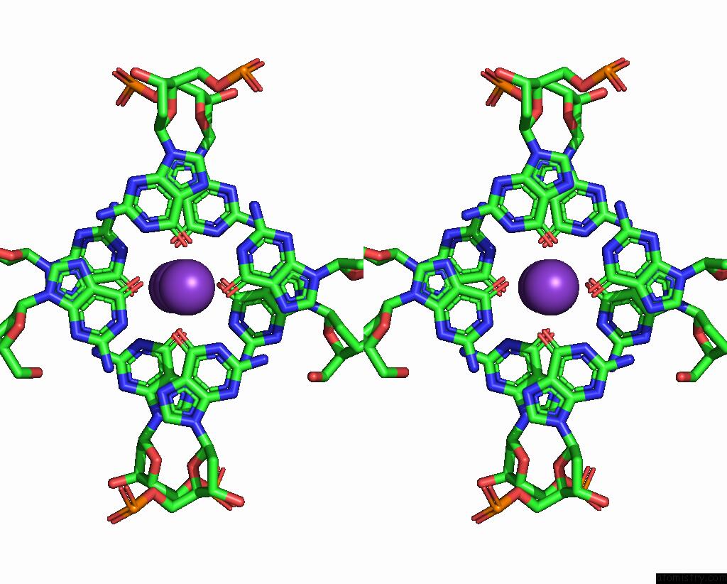
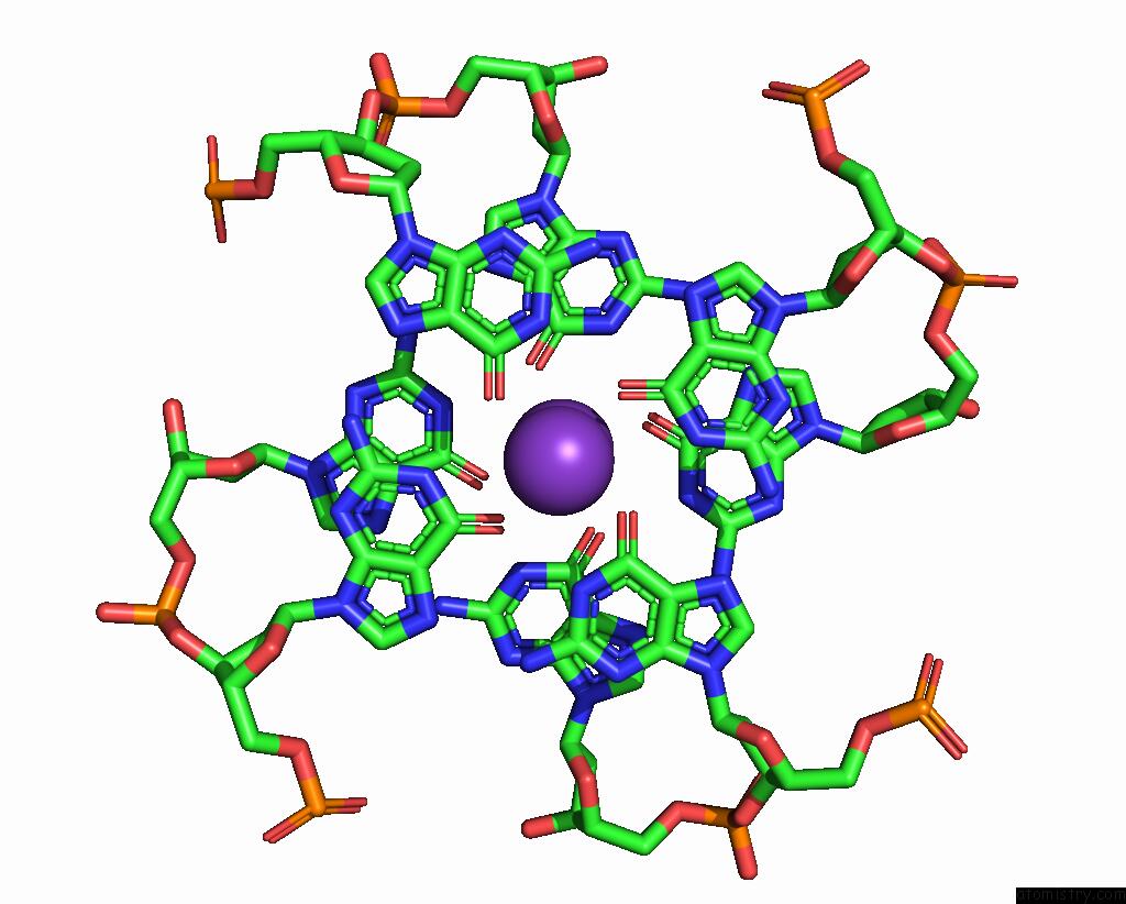
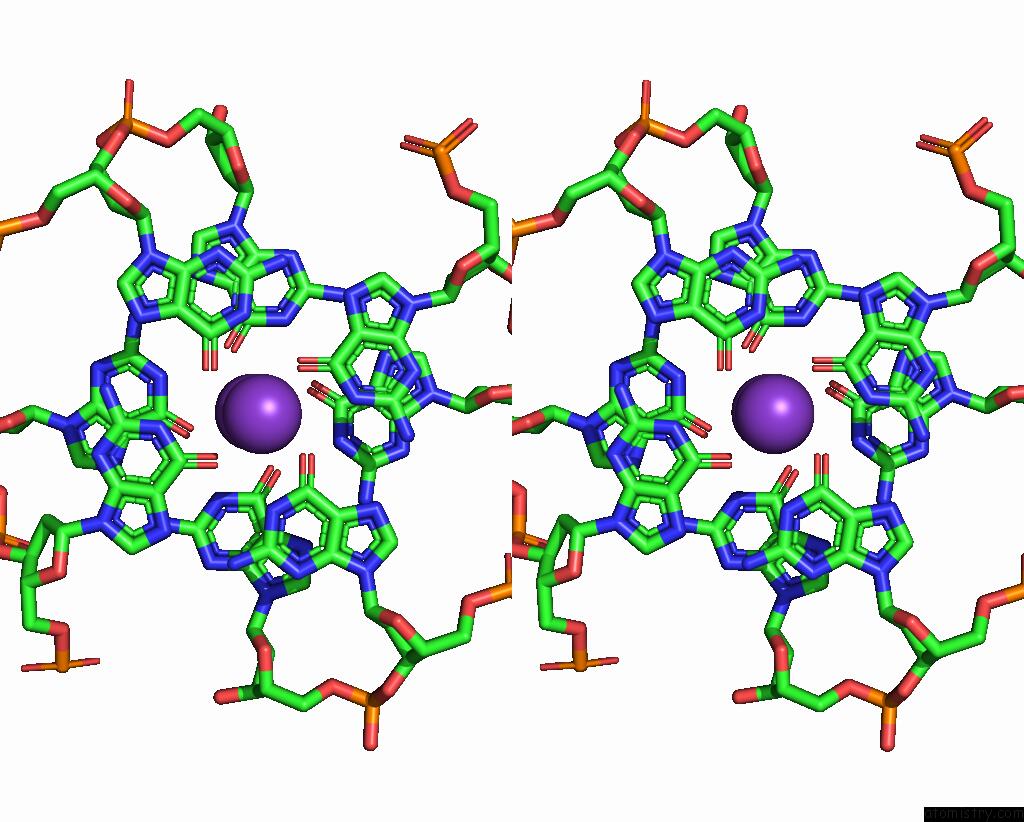
Potassium binding site 1 out of 5 in 7x7g
Go back to
Potassium binding site 1 out
of 5 in the Crystal Structure of A Dimeric Interlocked Parallel G-Quadruplex
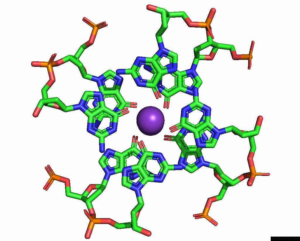
Mono view
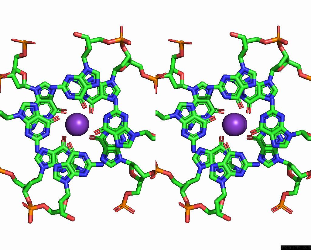
Stereo pair view
Mono view
Stereo pair view
A full contact list of Potassium with other atoms in the K binding
site number 1 of Crystal Structure of A Dimeric Interlocked Parallel G-Quadruplex within 5.0Å range:
|
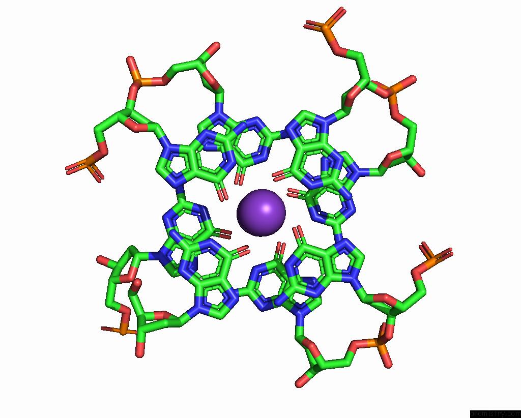
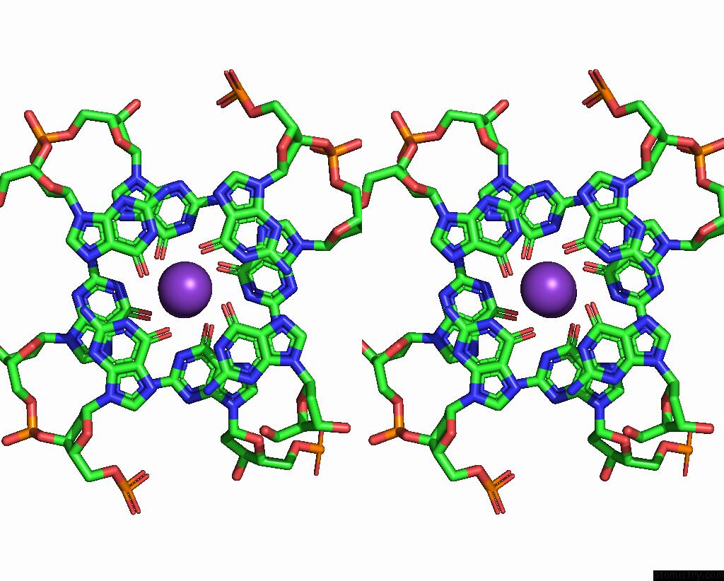
Potassium binding site 2 out of 5 in 7x7g
Go back to
Potassium binding site 2 out
of 5 in the Crystal Structure of A Dimeric Interlocked Parallel G-Quadruplex
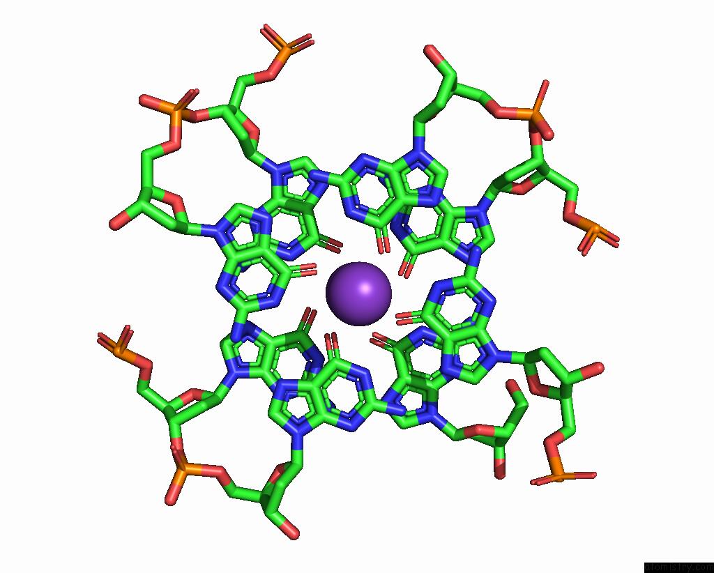
Mono view
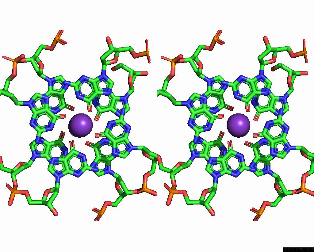
Stereo pair view
Mono view
Stereo pair view
A full contact list of Potassium with other atoms in the K binding
site number 2 of Crystal Structure of A Dimeric Interlocked Parallel G-Quadruplex within 5.0Å range:
|
Potassium binding site 3 out of 5 in 7x7g
Go back to
Potassium binding site 3 out
of 5 in the Crystal Structure of A Dimeric Interlocked Parallel G-Quadruplex
Mono view
Stereo pair view
Mono view
Stereo pair view
A full contact list of Potassium with other atoms in the K binding
site number 3 of Crystal Structure of A Dimeric Interlocked Parallel G-Quadruplex within 5.0Å range:
|
Potassium binding site 4 out of 5 in 7x7g
Go back to
Potassium binding site 4 out
of 5 in the Crystal Structure of A Dimeric Interlocked Parallel G-Quadruplex
Mono view
Stereo pair view
Mono view
Stereo pair view
A full contact list of Potassium with other atoms in the K binding
site number 4 of Crystal Structure of A Dimeric Interlocked Parallel G-Quadruplex within 5.0Å range:
|
Potassium binding site 5 out of 5 in 7x7g
Go back to
Potassium binding site 5 out
of 5 in the Crystal Structure of A Dimeric Interlocked Parallel G-Quadruplex
Mono view
Stereo pair view
Mono view
Stereo pair view
A full contact list of Potassium with other atoms in the K binding
site number 5 of Crystal Structure of A Dimeric Interlocked Parallel G-Quadruplex within 5.0Å range:
|
Reference:
K.H.Ngo,
C.W.Liew,
S.Lattmann,
F.R.Winnerdy,
A.T.Phan.
Crystal Structures of An Hiv-1 Integrase Aptamer: Formation of A Water-Mediated A.G.G.G.G Pentad in An Interlocked G-Quadruplex Biochem.Biophys.Res.Commun. 2022.
ISSN: ESSN 1090-2104
Page generated: Sat Aug 9 15:12:22 2025
ISSN: ESSN 1090-2104
Last articles
Mg in 3CP6Mg in 3CPJ
Mg in 3CPH
Mg in 3CON
Mg in 3CMV
Mg in 3COB
Mg in 3CNZ
Mg in 3CNX
Mg in 3CMX
Mg in 3CMW