Potassium »
PDB 7uut-7xx6 »
7vte »
Potassium in PDB 7vte: Uridine Bound Structure of Pseudouridine Kinase (Puki) From Escherichia Coli Strain B
Enzymatic activity of Uridine Bound Structure of Pseudouridine Kinase (Puki) From Escherichia Coli Strain B
All present enzymatic activity of Uridine Bound Structure of Pseudouridine Kinase (Puki) From Escherichia Coli Strain B:
2.7.1.83;
2.7.1.83;
Protein crystallography data
The structure of Uridine Bound Structure of Pseudouridine Kinase (Puki) From Escherichia Coli Strain B, PDB code: 7vte
was solved by
S.H.Kim,
S.Rhee,
with X-Ray Crystallography technique. A brief refinement statistics is given in the table below:
| Resolution Low / High (Å) | 36.89 / 2.15 |
| Space group | P 3 |
| Cell size a, b, c (Å), α, β, γ (°) | 185.689, 185.689, 50.754, 90, 90, 120 |
| R / Rfree (%) | 25 / 28.7 |
Potassium Binding Sites:
The binding sites of Potassium atom in the Uridine Bound Structure of Pseudouridine Kinase (Puki) From Escherichia Coli Strain B
(pdb code 7vte). This binding sites where shown within
5.0 Angstroms radius around Potassium atom.
In total 4 binding sites of Potassium where determined in the Uridine Bound Structure of Pseudouridine Kinase (Puki) From Escherichia Coli Strain B, PDB code: 7vte:
Jump to Potassium binding site number: 1; 2; 3; 4;
In total 4 binding sites of Potassium where determined in the Uridine Bound Structure of Pseudouridine Kinase (Puki) From Escherichia Coli Strain B, PDB code: 7vte:
Jump to Potassium binding site number: 1; 2; 3; 4;
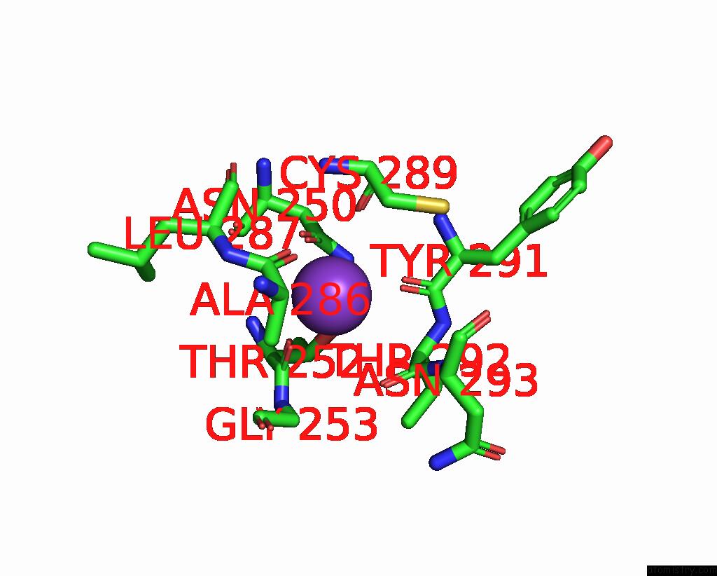
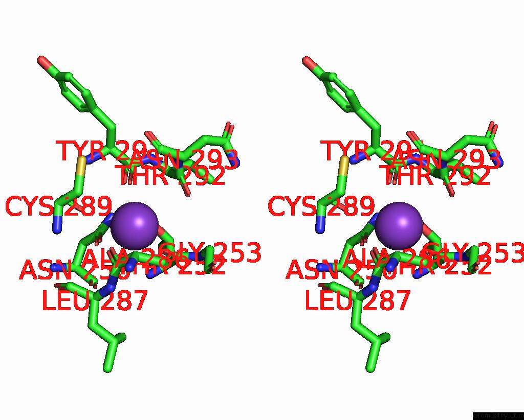
Potassium binding site 1 out of 4 in 7vte
Go back to
Potassium binding site 1 out
of 4 in the Uridine Bound Structure of Pseudouridine Kinase (Puki) From Escherichia Coli Strain B
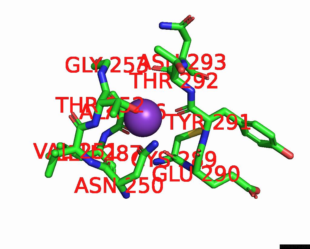
Mono view
Stereo pair view
Mono view
Stereo pair view
A full contact list of Potassium with other atoms in the K binding
site number 1 of Uridine Bound Structure of Pseudouridine Kinase (Puki) From Escherichia Coli Strain B within 5.0Å range:
|
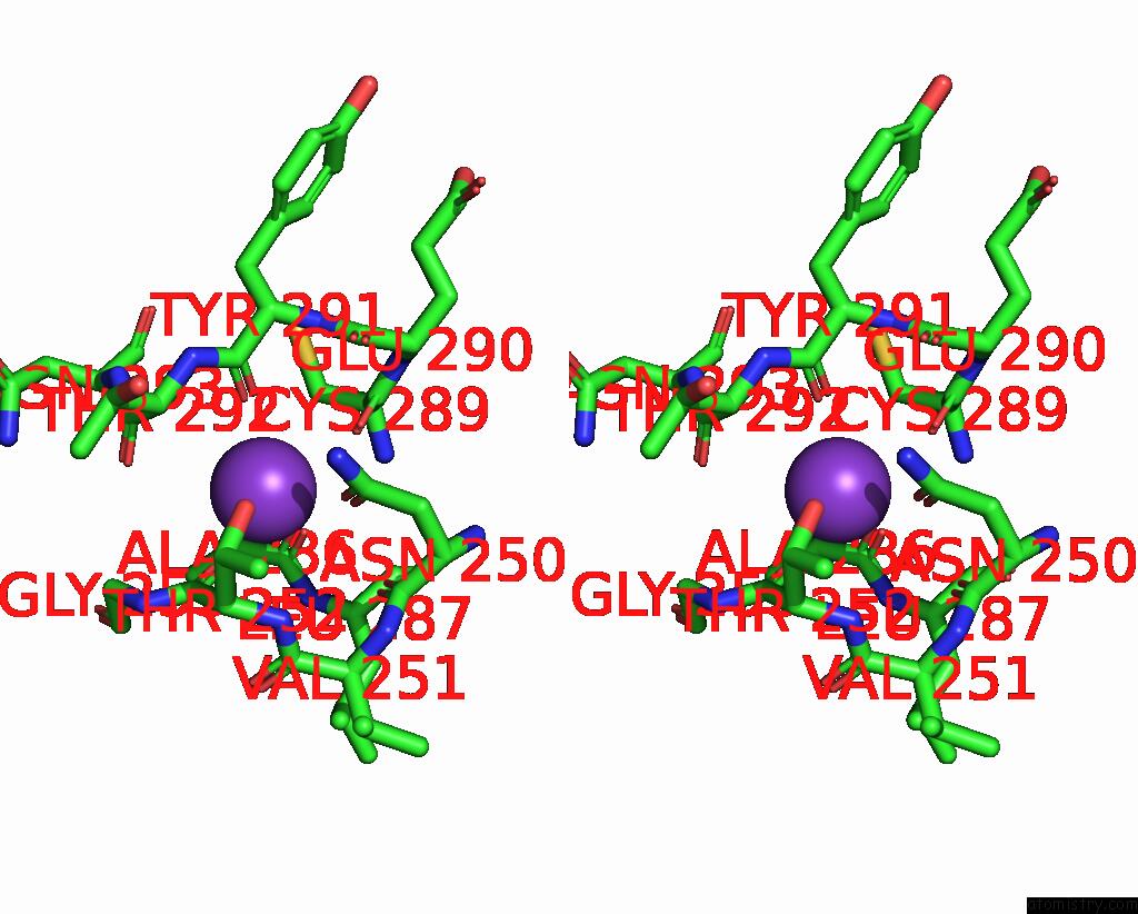
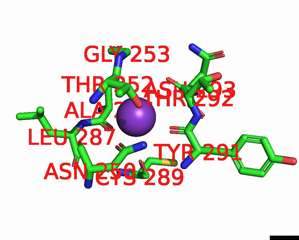
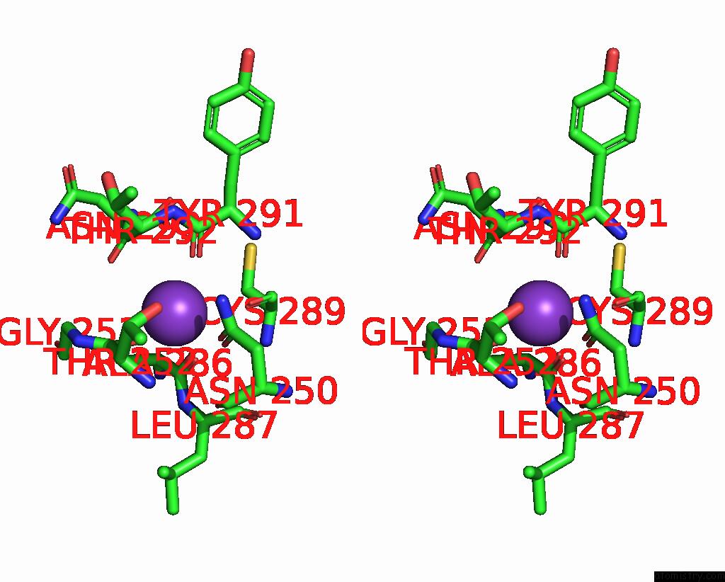
Potassium binding site 2 out of 4 in 7vte
Go back to
Potassium binding site 2 out
of 4 in the Uridine Bound Structure of Pseudouridine Kinase (Puki) From Escherichia Coli Strain B
Mono view
Stereo pair view
Mono view
Stereo pair view
A full contact list of Potassium with other atoms in the K binding
site number 2 of Uridine Bound Structure of Pseudouridine Kinase (Puki) From Escherichia Coli Strain B within 5.0Å range:
|
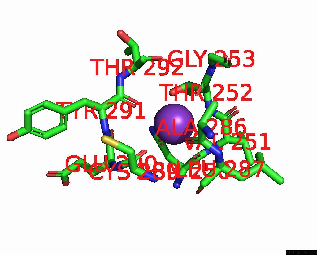
Potassium binding site 3 out of 4 in 7vte
Go back to
Potassium binding site 3 out
of 4 in the Uridine Bound Structure of Pseudouridine Kinase (Puki) From Escherichia Coli Strain B
Mono view
Stereo pair view
Mono view
Stereo pair view
A full contact list of Potassium with other atoms in the K binding
site number 3 of Uridine Bound Structure of Pseudouridine Kinase (Puki) From Escherichia Coli Strain B within 5.0Å range:
|
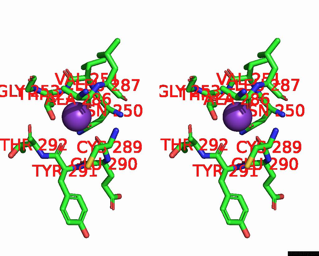
Potassium binding site 4 out of 4 in 7vte
Go back to
Potassium binding site 4 out
of 4 in the Uridine Bound Structure of Pseudouridine Kinase (Puki) From Escherichia Coli Strain B
Mono view
Stereo pair view
Mono view
Stereo pair view
A full contact list of Potassium with other atoms in the K binding
site number 4 of Uridine Bound Structure of Pseudouridine Kinase (Puki) From Escherichia Coli Strain B within 5.0Å range:
|
Reference:
S.H.Kim,
M.Kim,
D.Park,
S.Byun,
S.Rhee.
Substrate-Binding Loop Interactions with Pseudouridine Trigger Conformational Changes That Promote Catalytic Efficiency of Pseudouridine Kinase Puki. J.Biol.Chem. V. 298 01869 2022.
ISSN: ESSN 1083-351X
PubMed: 35346685
DOI: 10.1016/J.JBC.2022.101869
Page generated: Sat Aug 9 15:09:43 2025
ISSN: ESSN 1083-351X
PubMed: 35346685
DOI: 10.1016/J.JBC.2022.101869
Last articles
K in 9DITK in 9DJV
K in 9DIG
K in 9DID
K in 9DIC
K in 9DI8
K in 9DIB
K in 9DE5
K in 9D5W
K in 9D19