Potassium »
PDB 7uut-7xx6 »
7vlj »
Potassium in PDB 7vlj: Crystal Structure of Entamoeba Histolytica Serine Protease Inhibitor, Histopin, in the Cleaved Conformation
Protein crystallography data
The structure of Crystal Structure of Entamoeba Histolytica Serine Protease Inhibitor, Histopin, in the Cleaved Conformation, PDB code: 7vlj
was solved by
M.F.Ali,
S.Devi,
S.Gourinath,
with X-Ray Crystallography technique. A brief refinement statistics is given in the table below:
| Resolution Low / High (Å) | 45.64 / 1.83 |
| Space group | P 21 21 21 |
| Cell size a, b, c (Å), α, β, γ (°) | 39.13, 61.01, 137.253, 90, 90, 90 |
| R / Rfree (%) | 15.7 / 20.3 |
Potassium Binding Sites:
The binding sites of Potassium atom in the Crystal Structure of Entamoeba Histolytica Serine Protease Inhibitor, Histopin, in the Cleaved Conformation
(pdb code 7vlj). This binding sites where shown within
5.0 Angstroms radius around Potassium atom.
In total only one binding site of Potassium was determined in the Crystal Structure of Entamoeba Histolytica Serine Protease Inhibitor, Histopin, in the Cleaved Conformation, PDB code: 7vlj:
In total only one binding site of Potassium was determined in the Crystal Structure of Entamoeba Histolytica Serine Protease Inhibitor, Histopin, in the Cleaved Conformation, PDB code: 7vlj:
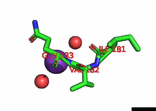
Potassium binding site 1 out of 1 in 7vlj
Go back to
Potassium binding site 1 out
of 1 in the Crystal Structure of Entamoeba Histolytica Serine Protease Inhibitor, Histopin, in the Cleaved Conformation
Mono view

Stereo pair view
Mono view
Stereo pair view
A full contact list of Potassium with other atoms in the K binding
site number 1 of Crystal Structure of Entamoeba Histolytica Serine Protease Inhibitor, Histopin, in the Cleaved Conformation within 5.0Å range:
|
Reference:
M.F.Ali,
S.Devi,
S.Gourinath.
Crystal Structure of Entamoeba Histolytica Serine Protease Inhibitor, Histopin, in the Cleaved Conformation To Be Published.
Page generated: Sat Aug 9 15:08:23 2025
Last articles
Mg in 3CPWMg in 3CZJ
Mg in 3D19
Mg in 3CZ4
Mg in 3CZ1
Mg in 3CZ0
Mg in 3CXO
Mg in 3CYZ
Mg in 3CYI
Mg in 3CX8