Potassium »
PDB 7pka-7qr1 »
7pv8 »
Potassium in PDB 7pv8: INLB392_T332E: T332E Variant of Listeria Monocytogenes Inlb (Internalin B) Residues 36-392
Protein crystallography data
The structure of INLB392_T332E: T332E Variant of Listeria Monocytogenes Inlb (Internalin B) Residues 36-392, PDB code: 7pv8
was solved by
C.Geerds,
H.H.Niemann,
with X-Ray Crystallography technique. A brief refinement statistics is given in the table below:
| Resolution Low / High (Å) | 16.15 / 2.05 |
| Space group | P 21 21 21 |
| Cell size a, b, c (Å), α, β, γ (°) | 44.58, 54.59, 226.72, 90, 90, 90 |
| R / Rfree (%) | 18.6 / 22.9 |
Potassium Binding Sites:
The binding sites of Potassium atom in the INLB392_T332E: T332E Variant of Listeria Monocytogenes Inlb (Internalin B) Residues 36-392
(pdb code 7pv8). This binding sites where shown within
5.0 Angstroms radius around Potassium atom.
In total only one binding site of Potassium was determined in the INLB392_T332E: T332E Variant of Listeria Monocytogenes Inlb (Internalin B) Residues 36-392, PDB code: 7pv8:
In total only one binding site of Potassium was determined in the INLB392_T332E: T332E Variant of Listeria Monocytogenes Inlb (Internalin B) Residues 36-392, PDB code: 7pv8:
Potassium binding site 1 out of 1 in 7pv8
Go back to
Potassium binding site 1 out
of 1 in the INLB392_T332E: T332E Variant of Listeria Monocytogenes Inlb (Internalin B) Residues 36-392
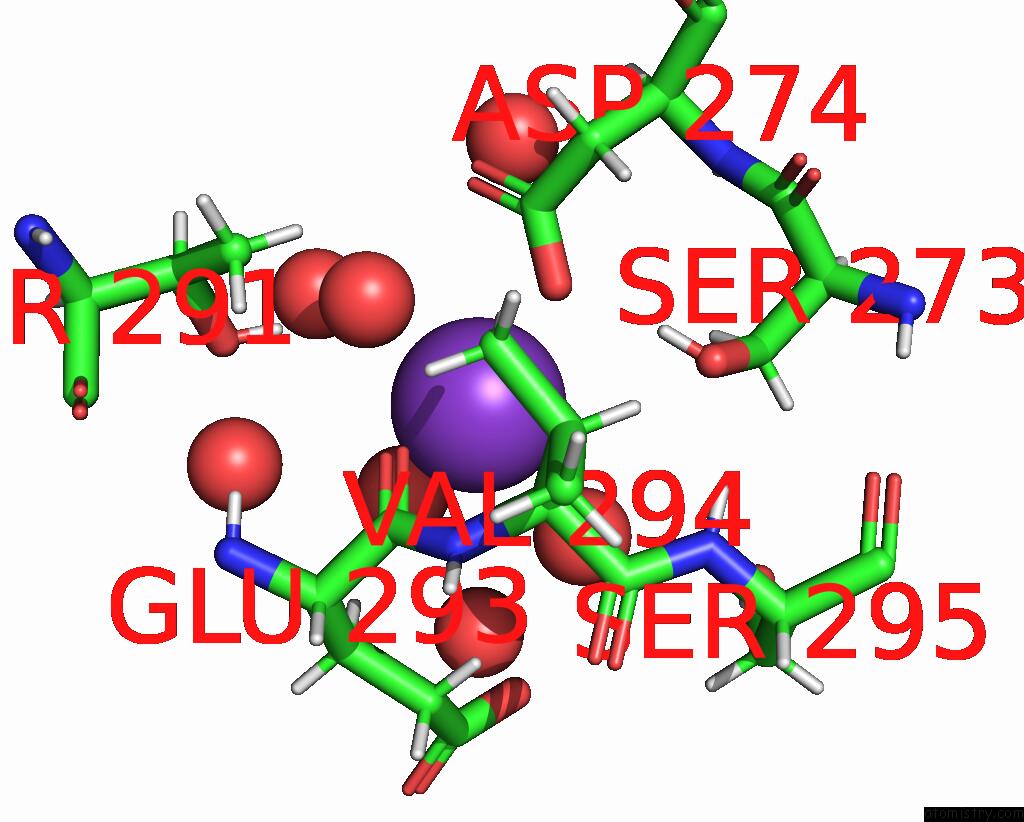
Mono view
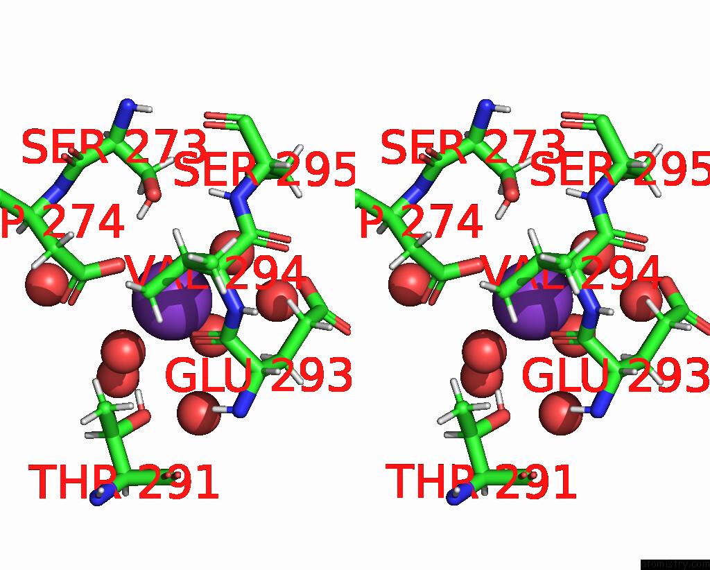
Stereo pair view
Mono view
Stereo pair view
A full contact list of Potassium with other atoms in the K binding
site number 1 of INLB392_T332E: T332E Variant of Listeria Monocytogenes Inlb (Internalin B) Residues 36-392 within 5.0Å range:
|
Reference:
C.Geerds,
W.M.Bleymuller,
T.Meyer,
C.Widmann,
H.H.Niemann.
A Recurring Packing Contact in Crystals of Inlb Pinpoints Functional Binding Sites in the Internalin Domain and the B Repeat. Acta Crystallogr D Struct V. 78 310 2022BIOL.
ISSN: ISSN 2059-7983
PubMed: 35234145
DOI: 10.1107/S2059798322000432
Page generated: Sat Aug 9 14:04:12 2025
ISSN: ISSN 2059-7983
PubMed: 35234145
DOI: 10.1107/S2059798322000432
Last articles
Mg in 4NXIMg in 4NX8
Mg in 4NV0
Mg in 4NWI
Mg in 4NX5
Mg in 4NV3
Mg in 4NW7
Mg in 4NRU
Mg in 4NST
Mg in 4NUA