Potassium »
PDB 7olt-7pji »
7p5t »
Potassium in PDB 7p5t: Structure of CYP142 From Mycobacterium Tuberculosis in Complex with Inhibitor MEK216
Enzymatic activity of Structure of CYP142 From Mycobacterium Tuberculosis in Complex with Inhibitor MEK216
All present enzymatic activity of Structure of CYP142 From Mycobacterium Tuberculosis in Complex with Inhibitor MEK216:
1.14.15.28;
1.14.15.28;
Protein crystallography data
The structure of Structure of CYP142 From Mycobacterium Tuberculosis in Complex with Inhibitor MEK216, PDB code: 7p5t
was solved by
M.Snee,
M.Kavanagh,
R.Tunnicliffe,
K.Mclean,
C.Levy,
A.Munro,
with X-Ray Crystallography technique. A brief refinement statistics is given in the table below:
| Resolution Low / High (Å) | 64.53 / 1.30 |
| Space group | P 21 21 21 |
| Cell size a, b, c (Å), α, β, γ (°) | 55.728, 65.717, 129.069, 90, 90, 90 |
| R / Rfree (%) | 13.7 / 16.2 |
Other elements in 7p5t:
The structure of Structure of CYP142 From Mycobacterium Tuberculosis in Complex with Inhibitor MEK216 also contains other interesting chemical elements:
| Iron | (Fe) | 1 atom |
| Bromine | (Br) | 3 atoms |
Potassium Binding Sites:
The binding sites of Potassium atom in the Structure of CYP142 From Mycobacterium Tuberculosis in Complex with Inhibitor MEK216
(pdb code 7p5t). This binding sites where shown within
5.0 Angstroms radius around Potassium atom.
In total 2 binding sites of Potassium where determined in the Structure of CYP142 From Mycobacterium Tuberculosis in Complex with Inhibitor MEK216, PDB code: 7p5t:
Jump to Potassium binding site number: 1; 2;
In total 2 binding sites of Potassium where determined in the Structure of CYP142 From Mycobacterium Tuberculosis in Complex with Inhibitor MEK216, PDB code: 7p5t:
Jump to Potassium binding site number: 1; 2;
Potassium binding site 1 out of 2 in 7p5t
Go back to
Potassium binding site 1 out
of 2 in the Structure of CYP142 From Mycobacterium Tuberculosis in Complex with Inhibitor MEK216
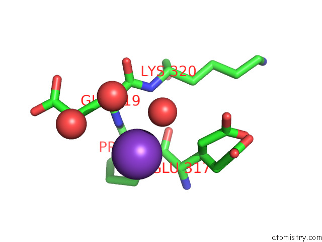
Mono view
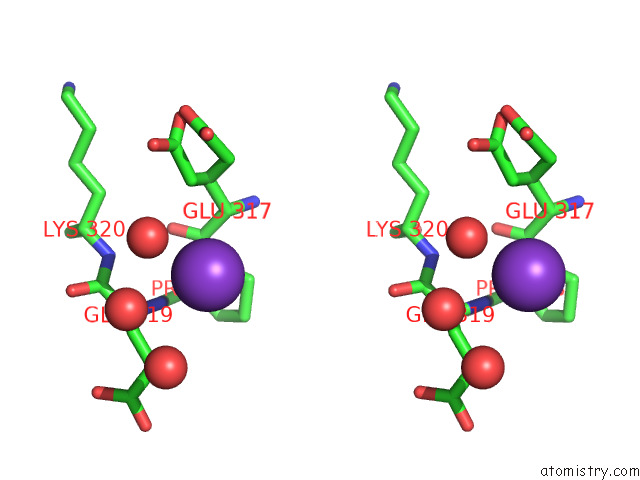
Stereo pair view
Mono view
Stereo pair view
A full contact list of Potassium with other atoms in the K binding
site number 1 of Structure of CYP142 From Mycobacterium Tuberculosis in Complex with Inhibitor MEK216 within 5.0Å range:
|
Potassium binding site 2 out of 2 in 7p5t
Go back to
Potassium binding site 2 out
of 2 in the Structure of CYP142 From Mycobacterium Tuberculosis in Complex with Inhibitor MEK216
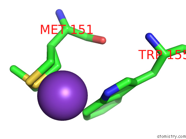
Mono view
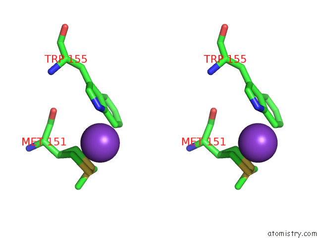
Stereo pair view
Mono view
Stereo pair view
A full contact list of Potassium with other atoms in the K binding
site number 2 of Structure of CYP142 From Mycobacterium Tuberculosis in Complex with Inhibitor MEK216 within 5.0Å range:
|
Reference:
M.Kavanagh,
A.Coyne.
Structure of CYP142 From Mycobacterium Tuberculosis in Complex with Inhibitor MEK216 To Be Published.
Page generated: Sat Aug 9 13:59:25 2025
Last articles
Mg in 2PRYMg in 2PRN
Mg in 2PPQ
Mg in 2PPB
Mg in 2PLY
Mg in 2PNQ
Mg in 2PP3
Mg in 2PP1
Mg in 2PMQ
Mg in 2PN3