Potassium »
PDB 7olt-7pji »
7ot4 »
Potassium in PDB 7ot4: Crystal Structure of Msra Variant C198C206 From Escherichia Coli, Oxidized
Enzymatic activity of Crystal Structure of Msra Variant C198C206 From Escherichia Coli, Oxidized
All present enzymatic activity of Crystal Structure of Msra Variant C198C206 From Escherichia Coli, Oxidized:
1.8.4.11;
1.8.4.11;
Protein crystallography data
The structure of Crystal Structure of Msra Variant C198C206 From Escherichia Coli, Oxidized, PDB code: 7ot4
was solved by
S.Napolitano,
R.Glockshuber,
with X-Ray Crystallography technique. A brief refinement statistics is given in the table below:
| Resolution Low / High (Å) | 43.48 / 2.19 |
| Space group | P 61 2 2 |
| Cell size a, b, c (Å), α, β, γ (°) | 128.21, 128.21, 118.34, 90, 90, 120 |
| R / Rfree (%) | 28.6 / 30.8 |
Potassium Binding Sites:
The binding sites of Potassium atom in the Crystal Structure of Msra Variant C198C206 From Escherichia Coli, Oxidized
(pdb code 7ot4). This binding sites where shown within
5.0 Angstroms radius around Potassium atom.
In total only one binding site of Potassium was determined in the Crystal Structure of Msra Variant C198C206 From Escherichia Coli, Oxidized, PDB code: 7ot4:
In total only one binding site of Potassium was determined in the Crystal Structure of Msra Variant C198C206 From Escherichia Coli, Oxidized, PDB code: 7ot4:
Potassium binding site 1 out of 1 in 7ot4
Go back to
Potassium binding site 1 out
of 1 in the Crystal Structure of Msra Variant C198C206 From Escherichia Coli, Oxidized
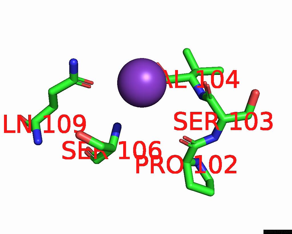
Mono view
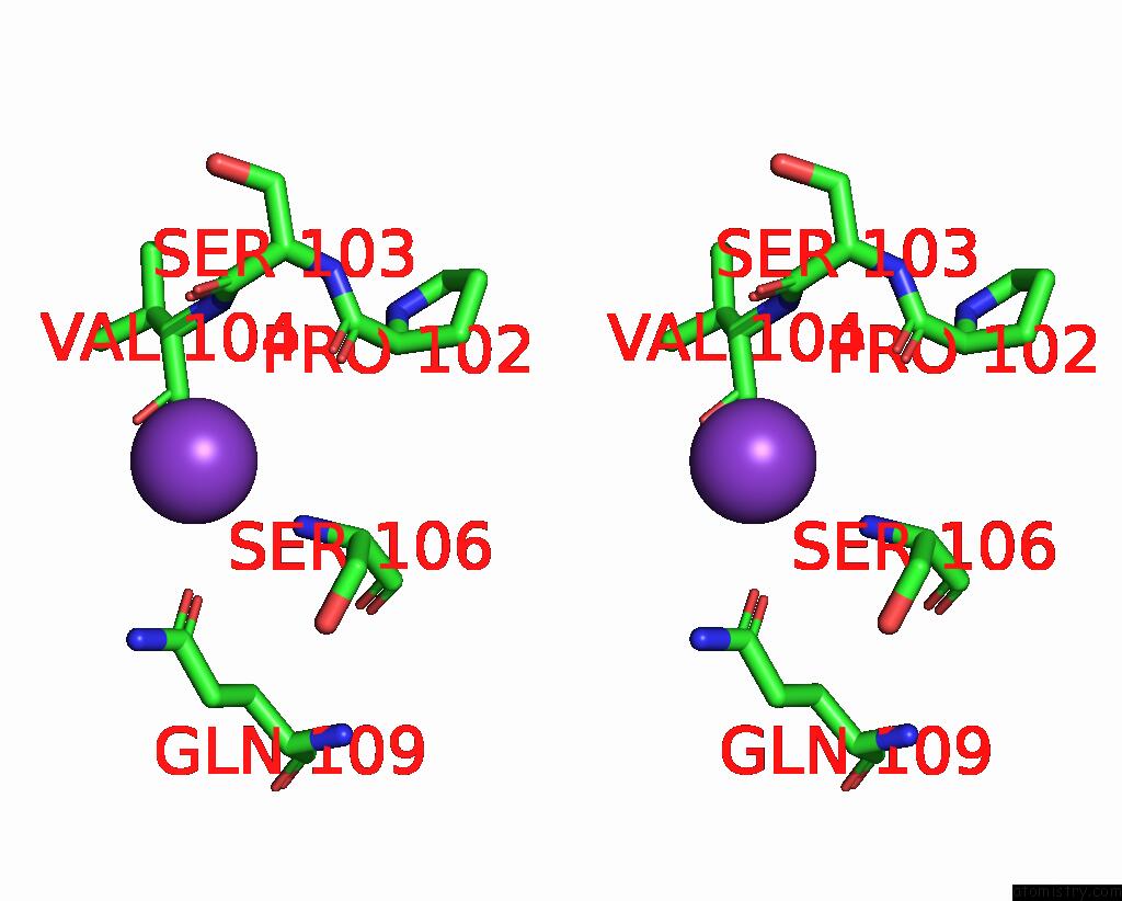
Stereo pair view
Mono view
Stereo pair view
A full contact list of Potassium with other atoms in the K binding
site number 1 of Crystal Structure of Msra Variant C198C206 From Escherichia Coli, Oxidized within 5.0Å range:
|
Reference:
S.Napolitano,
R.Glockshuber.
Exploring the Unique Mechanism of Methionine Sulphoxide Reduction By Escherichia Coli To Be Published.
Page generated: Sat Aug 9 13:53:32 2025
Last articles
Mg in 1MMNMg in 1MMG
Mg in 1MMD
Mg in 1MJI
Mg in 1MMA
Mg in 1MKJ
Mg in 1MJ5
Mg in 1MJN
Mg in 1MIY
Mg in 1MIW